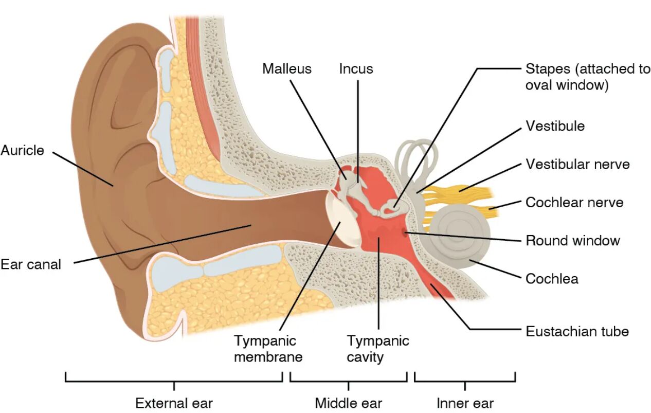
The ear is a remarkable organ that facilitates both hearing and balance, comprising the external, middle, and inner ear, each with distinct structures and functions. This image provides a detailed view of the auricle, tympanic membrane, ossicles, Eustachian tube, cochlea, and vestibule, illustrating their roles in auditory and equilibrium processes. This article offers an in-depth exploration of these anatomical components, enhancing understanding of their contributions to sensory perception and bodily stability.
Labeled Parts of the Ear
Auricle The auricle, or pinna, is the visible, cartilaginous outer part of the ear that collects sound waves and directs them into the ear canal. Its unique shape helps amplify and localize sound, enhancing auditory perception in different environments.
Ear canal The ear canal, or external auditory meatus, is a tube that extends from the auricle to the tympanic membrane, channeling sound waves inward. It is lined with cerumen-producing glands, which protect the canal and eardrum from debris and infections.
Tympanic membrane The tympanic membrane, or eardrum, is a thin, concave structure that separates the external ear from the middle ear, vibrating in response to sound waves. These vibrations are transmitted to the ossicles, initiating the process of hearing.
Ossicles The ossicles, consisting of the malleus, incus, and stapes, are three tiny bones in the middle ear that amplify and transfer sound vibrations from the tympanic membrane to the inner ear. Their precise movement ensures efficient sound conduction, with the stapes fitting into the oval window.
Eustachian tube The Eustachian tube connects the middle ear to the pharynx, equalizing air pressure on both sides of the tympanic membrane. It opens during swallowing or yawning, preventing pressure imbalances that could affect hearing.
Cochlea The cochlea is a spiral, fluid-filled structure in the inner ear responsible for converting sound vibrations into electrical signals for audition. Its internal hair cells detect specific frequencies, sending auditory information to the brain via the cochlear nerve.
Vestibule The vestibule is a central cavity in the inner ear that houses the utricle and saccule, contributing to the sense of balance and spatial orientation. It detects linear acceleration and head position, relaying this information to the vestibular nerve.
Anatomical Overview of the Ear
The ear’s structure is divided into three distinct regions, each contributing to its dual roles in hearing and balance. This organization ensures a seamless transition of sound and equilibrium signals from the external environment to the nervous system.
- External ear components: The auricle and ear canal capture and funnel sound waves, with the tympanic membrane serving as the gateway to the middle ear.
- Middle ear dynamics: The ossicles amplify vibrations, while the Eustachian tube maintains pressure equilibrium, protecting the delicate inner structures.
- Inner ear specialization: The cochlea processes auditory signals, and the vestibule handles balance, both encased in the temporal bone for protection.
- Connective pathways: The ear’s regions are linked by membranes and tubes, facilitating the transfer of mechanical and fluid-based signals.
- Protective features: Cerumen in the ear canal and the bony encasement of the inner ear safeguard against damage and infection.
Physiological Functions of the Ear
The ear’s anatomical structures work in concert to enable hearing and maintain equilibrium, adapting to a range of environmental conditions. These functions rely on the precise interaction of its components.
- Sound collection: The auricle gathers sound waves, directing them through the ear canal to vibrate the tympanic membrane.
- Vibration transmission: The ossicles amplify and transfer these vibrations to the cochlea, enhancing sound intensity for detection.
- Pressure regulation: The Eustachian tube equalizes middle ear pressure, ensuring the tympanic membrane functions optimally during altitude changes.
- Auditory processing: The cochlea’s hair cells convert vibrations into electrical impulses, which are sent to the brain for sound interpretation.
- Balance maintenance: The vestibule detects head movements and gravity, providing feedback to stabilize posture and coordination.
Developmental and Structural Dynamics
The ear’s anatomical structures develop through distinct embryonic stages, maturing to support hearing and balance by early childhood. This progression reflects both genetic programming and environmental influences.
- Embryonic origins: The auricle forms from hillocks of the first and second pharyngeal arches, while the inner ear derives from the otic placode.
- Ossicle formation: The ossicles develop from Meckel’s cartilage, migrating into the middle ear to establish the sound conduction chain.
- Cochlear development: The cochlea spirals during fetal growth, with hair cells differentiating to detect specific sound frequencies.
- Vestibular maturation: The vestibule’s sensory organs form early, enabling balance reflexes to emerge prenatally.
- Postnatal refinement: The Eustachian tube and ear canal continue to develop, adapting to respiratory and auditory demands.
Clinical Relevance and Ear Health
Understanding the ear’s anatomy is crucial for diagnosing and managing auditory and balance disorders. Clinical assessments often focus on these structures to identify issues and guide treatment.
- Otitis media: Inflammation of the middle ear, often involving the ossicles and Eustachian tube, can lead to hearing loss if untreated.
- Tinnitus: Persistent ringing may arise from cochlear damage or middle ear dysfunction, affecting quality of life.
- Vertigo: Vestibular disorders can cause dizziness, stemming from issues in the vestibule or its neural connections.
- Diagnostic tools: Audiometry and tympanometry evaluate hearing and middle ear function, while vestibular tests assess balance.
- Therapeutic options: Treatments range from antibiotics for infections to surgical interventions like tympanoplasty for eardrum repair.
In conclusion, the anatomical structures of the ear, spanning the external auricle to the inner cochlea and vestibule, form a sophisticated system for hearing and balance. This image underscores the intricate design that allows sound waves to be transformed into auditory experiences and head movements to be translated into equilibrium, offering a foundation for both health maintenance and clinical intervention. Exploring these components deepens appreciation for the ear’s dual sensory roles and the importance of preserving its function.

