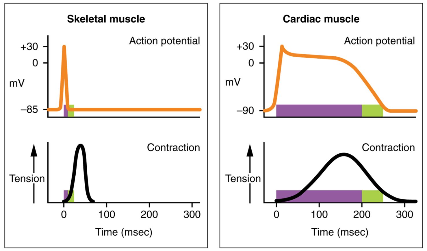
The action potential is a fundamental electrical event that drives muscle contraction, with distinct differences between heart and skeletal muscle that reflect their unique functions. This diagram compares the cardiac muscle action potential and skeletal muscle action potential, highlighting variations in duration, ion involvement, and refractory periods that support the heart’s rhythmic pumping versus skeletal muscle’s voluntary action. Exploring this image offers valuable insights into the electrophysiological adaptations of these muscle types.
Labelled Parts Explanation
- Cardiac muscle action potential The cardiac muscle action potential features a prolonged duration with a distinct plateau phase due to calcium ion influx, enabling sustained contraction. It includes an extended refractory period to prevent tetanic contraction, ensuring the heart completes its cycle before the next beat.
- Skeletal muscle action potential The skeletal muscle action potential is brief, relying on rapid sodium influx for depolarization and quick repolarization via potassium efflux, lacking a plateau phase. It has a short refractory period, allowing rapid successive contractions for voluntary movement.
Anatomical Overview of Muscle Action Potentials
The action potentials of cardiac and skeletal muscle are tailored to their specific physiological roles. This diagram illustrates the contrasting electrical profiles that support their functions.
- The cardiac muscle action potential sustains heart rhythm with a long plateau and refractory period.
- The skeletal muscle action potential enables quick, repetitive contractions for physical activity.
- The heart’s action potential depends heavily on calcium, while skeletal muscle relies on sodium.
- These differences reflect the involuntary versus voluntary nature of the muscles.
This comparison highlights their specialized adaptations.
Role of the Cardiac Muscle Action Potential
The cardiac muscle action potential is designed for continuous, rhythmic pumping. Its structure supports the heart’s demands.
- The cardiac muscle action potential includes a plateau phase lasting 200-300 ms, driven by calcium influx.
- This prolonged phase maintains contraction during ventricular ejection.
- The extended refractory period prevents re-excitation, ensuring complete relaxation.
- The process is regulated by the sinoatrial node for consistent rhythm.
This design is essential for sustaining circulation.
Function of the Skeletal Muscle Action Potential
The skeletal muscle action potential supports rapid, controlled movements. Its brevity suits voluntary action.
- The skeletal muscle action potential lasts 2-5 ms, with rapid sodium influx and repolarization.
- It lacks a plateau, allowing quick recovery for successive contractions.
- The short refractory period enables tetanic contractions under nervous control.
- This profile supports activities like running or lifting.
This structure aligns with skeletal muscle’s role.
Ionic Basis of Action Potentials
The action potentials depend on specific ion movements, differing between muscle types. These shifts drive electrical activity.
- The cardiac muscle action potential features a calcium influx during the plateau phase via L-type channels.
- The skeletal muscle action potential relies on sodium influx for depolarization and potassium for repolarization.
- Cardiac cells have a prolonged potassium efflux during the extended refractory period.
- These ionic differences reflect functional specialization.
This ionic interplay underpins muscle performance.
Physiological Importance of Action Potential Differences
The differences in action potentials optimize each muscle’s function. Their design supports distinct physiological needs.
- The cardiac muscle action potential’s plateau ensures adequate time for blood ejection.
- The skeletal muscle action potential’s brevity allows rapid, repeated contractions.
- The extended refractory period in cardiac cells prevents arrhythmias.
- The short refractory period in skeletal muscle supports sustained effort.
These adaptations are vital for their respective roles.
Comparison of Refractory Periods
The refractory periods vary significantly between cardiac and skeletal muscle. This distinction affects contraction patterns.
- The cardiac muscle action potential’s extended refractory period matches its contraction duration.
- The skeletal muscle action potential has a brief refractory period, enabling rapid firing.
- This prevents tetanus in the heart, while allowing it in skeletal muscle for strength.
- The difference reflects their rhythmic versus voluntary nature.
This feature ensures functional efficiency.
Clinical Relevance of Action Potential Variations
Understanding action potential differences aids in diagnosing muscle-related conditions. These profiles are key clinical markers.
- Prolongation of the cardiac muscle action potential’s plateau can indicate long QT syndrome.
- Abnormal skeletal muscle action potential firing may suggest myotonia or neuropathy.
- Electrocardiograms monitor cardiac changes, while electromyography assesses skeletal issues.
- Treatments target ion channels to correct these variations.
This knowledge supports effective medical interventions.
Conclusion
The action potential for heart muscle compared to skeletal muscle diagram provides a detailed comparison of the electrical events that drive their respective contractions. By exploring the cardiac muscle action potential with its long plateau and skeletal muscle action potential with its brief duration, one gains insight into how these profiles support the heart’s rhythm and skeletal muscle’s voluntary action. This understanding serves as a foundation for studying muscle physiology and addressing related health concerns, encouraging further exploration of the intricate electrophysiological designs that sustain bodily functions.

