The human lower limb is a marvel of evolutionary engineering, designed to support the weight of the entire body while providing the mobility necessary for walking, running, and jumping. At the core of this functionality is a complex arrangement of bones that form the leg’s structural framework. These bones are not merely static supports; they are dynamic tissues that interact with muscles, tendons, and ligaments to facilitate movement and protect vital structures. Understanding the anatomy of these bones is essential for medical professionals, athletes, and anyone interested in human physiology, as it provides insight into how our bodies maintain balance and withstand the physical stresses of daily life.
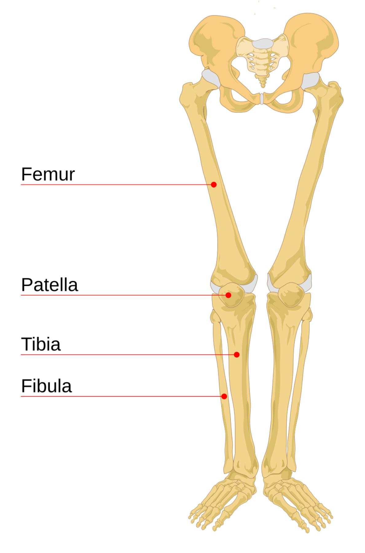
Femur: The femur is the thigh bone and stands as the longest, heaviest, and strongest bone in the human body. It articulates proximally with the acetabulum of the pelvis to form the hip joint and distally with the tibia and patella to form the knee joint.
Patella: The patella, commonly known as the kneecap, is a large sesamoid bone that sits in front of the knee joint within the quadriceps tendon. Its primary function is to increase the leverage of the quadriceps muscle and protect the anterior surface of the knee joint.
Tibia: The tibia, or shinbone, is the larger and stronger of the two bones in the lower leg and is located medially to the fibula. It serves as the main weight-bearing bone of the lower leg, connecting the knee to the ankle joint.
Fibula: The fibula is a slender bone located on the lateral side of the tibia, often referred to as the calf bone. While it does not bear significant body weight, it provides essential attachment points for muscles and helps stabilize the ankle joint through the lateral malleolus.
The Structural Framework of the Lower Limbs
The leg bones are a critical component of the skeletal system, providing the rigid levers required for locomotion. The arrangement begins at the hip, where the femur meets the pelvis, and extends down to the complex structure of the foot. This entire chain is optimized for upright posture, a defining characteristic of human biology. The bones of the leg are classified as long bones, meaning they have a shaft (diaphysis) and two expanded ends (epiphyses). This shape is ideal for resisting bending and compression forces while providing ample surface area for muscle attachment.
Beyond support, these bones house bone marrow, which is responsible for hematopoiesis, the production of blood cells. The density and mineralization of these bones are maintained through a constant process of remodeling, where old bone tissue is replaced by new tissue. This process is highly responsive to physical activity; weight-bearing exercises signal the body to increase bone density, making the legs stronger and more resilient over time.
Detailed Breakdown of Thigh and Knee Anatomy
The femur is arguably the most important bone in the lower limb due to its role in weight distribution. Its proximal end features a rounded head that fits into the pelvic socket, allowed for a wide range of motion at the hip. The neck of the femur, though strong, is a common site for fractures, especially in older populations with decreased bone density. Moving down the shaft, the femur ends in two large protrusions called condyles, which meet the tibia at the knee.
The knee joint is where the femur, tibia, and patella interact. It is one of the most complex joints in the body, relying on a sophisticated network of ligaments and menisci for stability. The patella plays a unique role here; by acting as a pulley, it allows the quadriceps to exert more force with less effort. This mechanical advantage is crucial for activities like climbing stairs or standing up from a seated position. The surfaces where these bones meet are covered in articular cartilage, a smooth tissue that minimizes friction and absorbs shock during movement.
The Lower Leg: Stability and Support
Below the knee, the lower leg consists of the tibia and the fibula. These two bones are connected by an interosseous membrane, a thick sheet of connective tissue that provides stability and serves as an attachment site for various muscles. The tibia is the workhorse of the lower leg, bearing the vast majority of the weight transferred from the femur. Its prominent anterior ridge is what we commonly feel as the “shin.”
- The medial malleolus of the tibia forms the inner prominence of the ankle.
- The lateral malleolus of the fibula forms the outer prominence of the ankle.
- The proximal tibiofibular joint allows for slight movement to accommodate ankle rotation.
- Muscles of the calf, such as the gastrocnemius, attach to the posterior surfaces of these bones.
While the fibula is much thinner than the tibia, its presence is vital for the integrity of the ankle. It acts as a lateral brace, preventing the ankle from rolling excessively. Furthermore, many of the muscles that move the foot and toes originate on the fibula, highlighting its importance in fine motor control and balance during movement on uneven terrain.
Common Injuries and Clinical Significance
Given the high loads they carry, the bones of the leg are susceptible to various injuries. Fractures of the femur usually require high-impact trauma, such as a motor vehicle accident, due to the bone’s immense strength. In contrast, stress fractures of the tibia are common among long-distance runners who increase their mileage too quickly. These tiny cracks occur when the muscles become fatigued and can no longer absorb the shock of impact, transferring the stress directly to the bone.
The knee and ankle joints are also frequent sites of pathology. Patellar tracking disorder occurs when the kneecap does not slide properly within the femoral groove, leading to pain and inflammation. Ankle fractures often involve the malleoli of the tibia or fibula and can significantly impair mobility. Understanding the specific landmarks on these bones helps clinicians diagnose injuries through physical examination and imaging techniques like X-rays and MRI scans.
Maintaining Bone Health in the Legs
Protecting the health of the leg bones is a lifelong endeavor that involves nutrition, exercise, and safety. Calcium and Vitamin D are the primary building blocks for bone tissue; without them, bones become brittle and prone to conditions like osteoporosis. Regular weight-bearing exercise, such as walking or resistance training, is equally important as it stimulates osteoblast activity, the cells responsible for building bone.
Additionally, wearing supportive footwear and using proper form during athletic activities can prevent acute injuries and chronic wear-and-tear. As we age, maintaining balance through proprioceptive training can reduce the risk of falls, which are a leading cause of hip and leg fractures in the elderly. By appreciating the intricate design of the human leg bones, we can better care for these structures that carry us through life.

