The biological group known as the Class Trematoda represents one of the most clinically significant lineages within the Phylum Platyhelminthes. Commonly referred to as flukes, these organisms have evolved into highly specialized endoparasites that inhabit the internal organs of vertebrates, including humans and various livestock. Among the many families within this class, the Fasciolidae stand out due to their considerable size and the severe pathological impact they have on the hepatic systems of their hosts. This group includes well-known species such as the common liver fluke and the giant liver fluke. These parasites are characterized by their leaf-like morphology, complex life cycles involving aquatic snails as intermediate hosts, and their ability to cause chronic disease that leads to significant economic losses in the global agricultural sector. Understanding the morphological distinctions and the ecological behavior of these flukes is paramount for effective diagnosis, treatment, and epidemiological control.
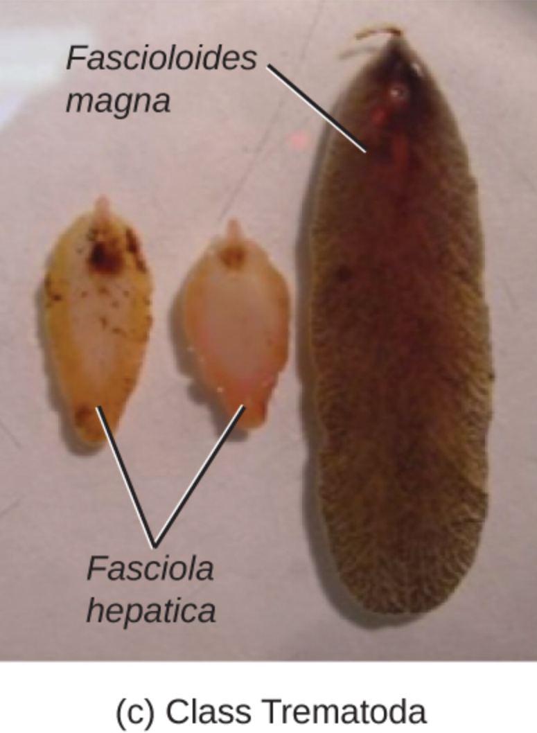
Fascioloides magna: This label points to the large, dark specimen on the right side of the image, commonly known as the giant liver fluke. It is significantly larger than its relatives, often reaching lengths of up to 10 centimeters, and primarily targets the liver tissue of wild ruminants like deer and elk.
Fasciola hepatica: This label identifies the two smaller, lighter-colored specimens on the left, which are known as common liver flukes. These parasites are a major cause of fascioliasis in sheep, cattle, and humans, characterized by their flattened, leaf-like body and a distinct cone-shaped anterior end.
Class Trematoda: This taxonomic designation at the bottom of the image groups these specimens into the class of parasitic flatworms known as flukes. Members of this class possess suckers for attachment and usually exhibit a complex life cycle requiring at least two different host species to reach maturity.
General Characteristics of the Class Trematoda
The Class Trematoda consists of thousands of species, almost all of which are obligate parasites. They are typically divided into two subclasses: Monogenea (which are mostly ectoparasites) and Digenea (the endoparasitic flukes). The specimens shown in the image belong to the Digenea subclass. These organisms are distinguished by their dorsoventrally flattened bodies, which facilitate their movement through the narrow bile ducts and tissues of their hosts. Unlike the free-living flatworms, trematodes have lost many of their external sensory organs but have gained specialized structures for attachment and nutrient absorption.
Structurally, trematodes possess two main suckers: an oral sucker surrounding the mouth at the anterior end and a ventral sucker, or acetabulum, located on the mid-ventral surface. These suckers allow the fluke to maintain its position within the host despite the flow of fluids. Their digestive system is incomplete, consisting of a pharynx and a bifid intestine (two cecae) that branch throughout the body. Because they lack a circulatory system, the branched gut plays a vital role in distributing nutrients. Most trematodes are hermaphroditic, containing both male and female reproductive systems within a single individual, allowing for self-fertilization or cross-fertilization depending on the availability of mates.
The Biology of Fasciola hepatica
Fasciola hepatica is perhaps the most famous member of the fluke family. It is a cosmopolitan parasite, found in temperate regions across the globe where sheep and cattle are raised. The adult fluke typically measures between 20 and 30 millimeters in length and about 10 millimeters in width. One of its most identifying features is the “cephalic cone,” a distinct projection at the front of the body where the oral sucker is located. The body behind this cone widens into “shoulders” before tapering toward the posterior end.
In terms of pathology, Fasciola hepatica is the primary agent of fascioliasis. In humans, the infection is often acquired by consuming raw aquatic plants, such as watercress, that have been contaminated with the encysted larval stage. Once ingested, the young flukes burrow through the intestinal wall, migrate across the peritoneal cavity, and eventually penetrate the liver capsule. Their migration through the liver tissue causes mechanical damage and triggers an inflammatory response, leading to fibrosis and bile duct obstruction. This disease can be chronic, lasting for years if left untreated, resulting in anemia, jaundice, and general wasting.
Comparing Fascioloides magna to Its Relatives
The specimen labeled Fascioloides magna provides a stark contrast to Fasciola hepatica in terms of scale. While Fasciola is measured in millimeters, Fascioloides can be several centimeters long and quite thick. Its body is more oval-shaped and lacks the distinct cephalic cone seen in its smaller cousin. This species is native to North America but was introduced to Europe in the 19th century through the importation of game animals. Its primary hosts are members of the deer family (Cervidae), where the fluke typically resides in fibrous cysts within the liver parenchyma.
The impact of Fascioloides magna varies significantly depending on the host species. In its natural deer hosts, the parasite and the host have co-evolved such that the infection is rarely fatal. However, when the fluke infects “dead-end” or “aberrant” hosts like cattle or sheep, the results are much more severe. In cattle, the parasite is usually encapsulated in thick-walled cysts, which prevents the eggs from reaching the bile ducts and exiting the body, effectively stopping the life cycle. In sheep and goats, the fluke continues to migrate through the liver without ever becoming encapsulated, causing massive tissue destruction and often leading to the death of the host before the parasite can even mature.
The Complex Life Cycle of Liver Flukes
The life cycle of these trematodes is a marvel of biological complexity, requiring a series of environmental transitions and host changes. It begins when adult flukes in the liver of a definitive host (a mammal) produce eggs that pass into the bile ducts and are eventually excreted in the feces. For the cycle to continue, these eggs must reach a freshwater environment. Under the right conditions of moisture and temperature, the egg hatches to release a ciliated, free-swimming larva called a miracidium.
- Intermediate Host Phase: The miracidium has a very short window (about 24 hours) to find and penetrate a specific type of freshwater snail, usually from the family Lymnaeidae. Inside the snail, the parasite undergoes several stages of asexual reproduction, transforming from a sporocyst into rediae, and eventually into numerous cercariae.
- Free-living Phase: The cercariae emerge from the snail and swim to aquatic vegetation. There, they lose their tails and encyst, becoming metacercariae. This stage is highly resistant to environmental stressors and can survive for months in damp conditions.
- Definitive Host Phase: When a mammal grazes on the contaminated vegetation, the metacercariae excyst in the duodenum, and the cycle begins anew.
Clinical Significance and Veterinary Management
The economic impact of these flukes cannot be overstated. In livestock, infections result in reduced milk production, poor weight gain, and the condemnation of livers at slaughterhouses. In many parts of the world, liver fluke control is a routine part of farm management. This involves a combination of tactical drenching with anthelmintic drugs, such as triclabendazole, and pasture management techniques designed to reduce the snail population. Fencing off boggy or poorly drained areas of fields where snails thrive is often a highly effective non-chemical strategy.
In human medicine, diagnosis is typically achieved through the detection of eggs in stool samples or via serological tests that detect antibodies against fluke antigens. Imaging techniques like ultrasound or CT scans can also reveal the presence of adult flukes in the bile ducts or the characteristic tracks left by migrating larvae in the liver tissue. While human fascioliasis is relatively rare in developed nations, it remains a significant public health challenge in parts of South America, Egypt, and Southeast Asia, where cultural dietary habits and proximity to livestock increase the risk of transmission.
Conclusion: Evolutionary Excellence and Parasitic Persistence
The specimens of Fasciola hepatica and Fascioloides magna shown in the image represent a pinnacle of parasitic adaptation. Through their sophisticated life cycles and specialized anatomy, they have secured a place in almost every corner of the world where water and mammals meet. As our climate changes and global trade in livestock continues, the range of these parasites may shift, posing new challenges for both veterinary and human health professionals. Continued research into the molecular biology of the Class Trematoda is essential for developing new diagnostic tools and vaccines, ensuring that we can effectively manage the threat posed by these ancient and resilient organisms.
Key Points Summary
- Trematodes are flatworms with complex life cycles and specialized suckers.
- Fasciola hepatica is a major human and livestock pathogen causing fascioliasis.
- Fascioloides magna is much larger and particularly dangerous to sheep and cattle.
- Freshwater snails are critical intermediate hosts for the transmission of liver flukes.
- Control requires a multifaceted approach involving medication and land management.

