Roundworms, belonging to the phylum Nematoda, represent one of the most diverse and medically significant groups of organisms on Earth. These elongated, unsegmented creatures thrive in countless environments, from soil to the human body, where certain species like Enterobius vermicularis, commonly known as the pinworm, cause widespread infections particularly among children. This microscopic view reveals the delicate yet resilient anatomy of a nematode, offering valuable insights into its structure and the reasons it remains a common concern in clinical practice worldwide. Understanding these organisms helps medical professionals and the public alike recognize symptoms, implement prevention strategies, and appreciate the intricate biology that allows nematodes to persist as both free-living and parasitic entities.
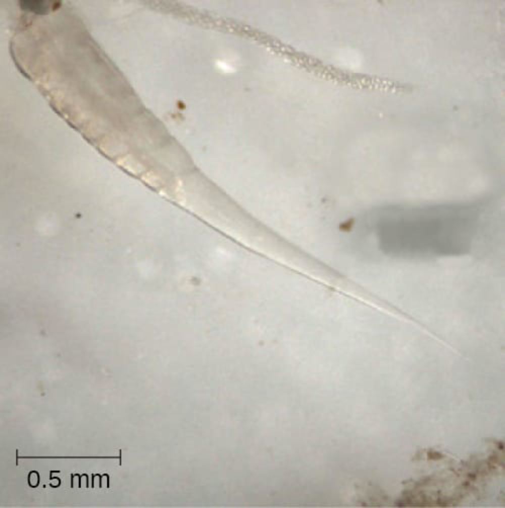
0.5 mm scale bar This measurement indicator provides essential context for appreciating the microscopic size of the specimen. It demonstrates that the entire length of the visible nematode is just over a few millimeters, highlighting how these parasites can easily go unnoticed without magnification while still being capable of causing significant discomfort in hosts.
The image displays a translucent, elongated organism characteristic of nematodes, with a tapered posterior end and a more robust anterior region. No additional textual labels appear directly on the micrograph besides the scale, but the visual features align with the typical morphology of Enterobius vermicularis, a common human pinworm.
Introduction to Phylum Nematoda
Nematodes, or roundworms, constitute a vast phylum within the animal kingdom, estimated to include over 25,000 described species with many more yet to be discovered. Their cylindrical, thread-like bodies lack segmentation, distinguishing them from annelids, while a tough yet flexible cuticle protects them from environmental stresses and host immune responses. In medical contexts, nematodes are responsible for some of the most prevalent parasitic infections globally, affecting billions of people, particularly in regions with limited sanitation. The specimen shown, identified as Enterobius vermicularis, exemplifies a nematode adapted specifically for human intestinal habitation.
Biology and Morphology of Enterobius vermicularis
Enterobius vermicularis, the pinworm, is a small white nematode that primarily infects the large intestine of humans. Adult females measure approximately 8-13 mm in length, while males are smaller at 2-5 mm. The organism in the image exhibits the classic tapered tail that gives pinworms their name, along with a smooth, transparent body wall that allows internal structures to be partially visible under microscopy. This transparency is due to the thin cuticle and lack of heavy pigmentation, an adaptation that aids in their survival within the host’s gut environment.
Pinworms possess a complete digestive tract, including a mouth with three lips and a simple esophagus leading to an intestine. They do not have a circulatory or respiratory system, relying instead on diffusion for gas exchange across their body surface. Reproduction is sexual, with females laying thousands of eggs per day, usually at night around the perianal region, which leads to intense itching and facilitates transmission through the fecal-oral route.
Life Cycle and Transmission of Pinworms
The life cycle of Enterobius vermicularis is direct and does not require an intermediate host, making it highly contagious in close-contact settings such as households, schools, and daycare centers. Eggs are ingested, hatch in the small intestine, and larvae mature in the large intestine within a few weeks. Gravid females migrate to the anus to deposit eggs, which become infective within hours and can survive on surfaces for up to three weeks. This rapid cycle explains why reinfection is common even after treatment.
Transmission occurs primarily through contaminated hands, bedding, clothing, or dust containing eggs. Children are most frequently affected due to behaviors like thumb-sucking or poor hand hygiene. In the provided micrograph, the slender, pointed posterior end is typical of female pinworms, which use this shape to anchor while laying eggs externally.
Clinical Features and Symptoms of Pinworm Infection
Enterobiasis, the infection caused by pinworms, is often mild but can cause significant discomfort. The hallmark symptom is nocturnal perianal pruritus, or intense itching around the anus at night, resulting from the female worm’s egg-laying activity. This itching can lead to sleep disturbances, irritability, and secondary bacterial skin infections from scratching. In some cases, especially with heavy infestations, abdominal pain, nausea, or even appendicitis-like symptoms may occur if worms migrate into the appendix.
Diagnosis is typically confirmed using the “tape test,” where adhesive tape is applied to the perianal area in the morning and examined microscopically for characteristic eggs, which are oval and flattened on one side. Adult worms may also be visible in stool or on bedding. The image provided illustrates the adult worm’s appearance, which is crucial for laboratory identification during medical training.
Diagnosis and Laboratory Identification
Microscopic examination remains the gold standard for identifying nematodes like Enterobius vermicularis. The specimen in the micrograph shows the worm’s elongated body, tapered tail, and translucent appearance, features that distinguish it from other intestinal parasites such as hookworms or Ascaris. In clinical labs, worms or eggs are often viewed at similar magnifications, with the 0.5 mm scale helping technicians gauge size accurately.
Advanced techniques including PCR can detect pinworm DNA, but they are rarely needed due to the simplicity of visual diagnosis. Proper sample collection and awareness of the worm’s morphology prevent misidentification with similar-looking artifacts or other nematodes.
Treatment and Management of Enterobiasis
Effective treatment for pinworm infection involves antiparasitic medications such as mebendazole, albendazole, or pyrantel pamoate, usually administered in a single dose with a repeat after two weeks to address newly hatched larvae. All household members should be treated simultaneously to break the transmission cycle. Supportive measures include thorough hand washing, laundering bedding in hot water, and trimming fingernails to reduce egg spread.
Prevention focuses on hygiene education, particularly in pediatric populations. Public health campaigns emphasize that pinworms, while bothersome, are not associated with serious long-term complications in most cases and respond well to therapy. The visual reference of the nematode in this image serves as an educational tool for healthcare providers to recognize and manage such infections confidently.
Epidemiology and Global Impact of Nematode Infections
Phylum Nematoda includes both free-living and parasitic species, with parasitic forms causing diseases ranging from mild enterobiasis to severe conditions like lymphatic filariasis or river blindness caused by other nematodes. Enterobius vermicularis has a cosmopolitan distribution, infecting up to 50% of children in some communities despite being less common in adults. Improved sanitation has reduced overall nematode burdens in developed regions, yet pinworms persist due to their efficient transmission method.
Globally, soil-transmitted helminths, a group that includes several nematodes, affect over 1.5 billion people according to health organizations. Understanding the basic morphology shown in the micrograph helps illustrate why these parasites are so successful: their simple body plan, protective cuticle, and reproductive efficiency enable them to thrive in diverse conditions.
Comparative Anatomy Within Phylum Nematoda
While Enterobius vermicularis is relatively small and harmless compared to larger parasites like Ascaris lumbricoides, which can grow up to 35 cm, all nematodes share fundamental traits: a pseudocoelom, longitudinal muscles only (resulting in a characteristic thrashing motion), and a collagenous cuticle that molts during growth. The image highlights the smooth, cylindrical form without obvious segmentation or appendages, features that are consistent across the phylum.
Other medically important nematodes include hookworms (Necator and Ancylostoma), which attach to the intestinal wall and cause anemia, and Trichinella, responsible for trichinellosis from undercooked meat. Each has unique adaptations visible under microscopy, but the basic roundworm architecture remains the same as seen here.
Educational Value of Microscopic Images in Medical Training
Micrographs like the one featured play a vital role in parasitology education. By visualizing the actual size via the scale bar and observing fine details of the worm’s body, students and practitioners develop pattern recognition skills essential for accurate diagnosis. Such images bridge theoretical knowledge with practical laboratory work, emphasizing the importance of careful microscopic technique when examining stool samples or perianal swabs.
In modern medical curricula, digital resources allow widespread access to high-quality images of nematodes, enhancing understanding of their life cycles, pathology, and control measures. This particular image of Enterobius vermicularis serves as a clear example for identifying one of the most common yet often underappreciated parasitic infections.
In conclusion, the study of phylum Nematoda, exemplified by Enterobius vermicularis, underscores the delicate balance between human health and parasitic organisms. Through continued research, education, and simple hygiene practices, the impact of these roundworms can be effectively minimized, ensuring better quality of life especially for vulnerable populations.

