Paramecium stands as one of the most iconic and extensively studied single-celled eukaryotes, serving as a cornerstone model organism in cell biology, genetics, and ciliary research for over a century. This slipper-shaped ciliate, belonging to the supergroup Chromalveolata, demonstrates complex behaviors and sophisticated cellular machinery despite its unicellular nature, offering profound insights into processes conserved across eukaryotes, including humans. From coordinated ciliary beating that powers motility and feeding to nuclear dimorphism and osmoregulation, Paramecium provides an accessible system for exploring fundamental biological principles with direct relevance to human health, particularly in understanding ciliopathies and cellular signaling.
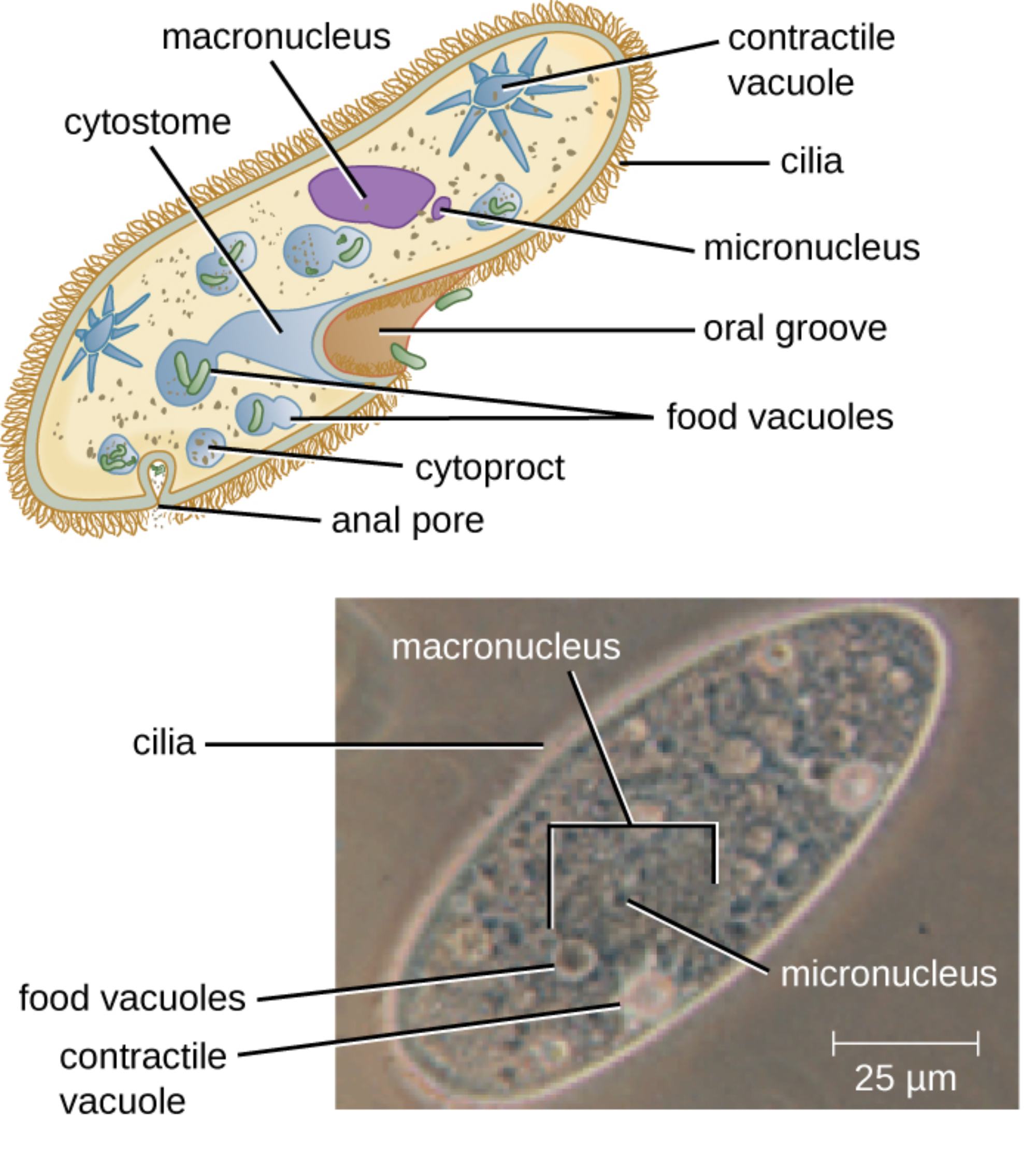
Macronucleus is the large, polyploid nucleus visible in both the diagram and microscopic view. It controls everyday cellular functions such as metabolism, growth, and gene expression through its abundant DNA copies, allowing high levels of transcriptional activity required for the organism’s active lifestyle.
Micronucleus is the small, diploid nucleus located near the macronucleus. It primarily serves as the germline nucleus, storing genetic material for sexual reproduction via conjugation and maintaining genomic integrity across generations.
Contractile vacuole is the star-shaped organelle responsible for osmoregulation. It collects excess water from the cytoplasm through radiating canals and periodically contracts to expel the fluid through a pore, preventing the cell from bursting in its hypotonic freshwater environment.
Cilia are the numerous hair-like projections covering the entire surface of the cell. These motile organelles beat in coordinated metachronal waves to propel the organism through water and generate currents that direct food particles toward the oral groove.
Cytostome is the mouth-like opening or cell mouth located in the oral groove. It serves as the entry point for food particles, which are swept in by ciliary action and engulfed into food vacuoles for digestion.
Oral groove is the funnel-shaped depression on one side of the cell that leads to the cytostome. It channels water currents carrying bacteria and organic debris toward the feeding apparatus, enhancing feeding efficiency.
Food vacuoles are membrane-bound sacs containing ingested food particles at various stages of digestion. They circulate through the cytoplasm, fusing with lysosomes to break down nutrients before waste is expelled.
Cytoproct is the anal pore or cell anus located posteriorly. It serves as the site for egestion, where indigestible residues from food vacuoles are expelled from the cell.
Anal pore refers to the same structure as the cytoproct, the posterior opening through which waste materials are discharged after digestion is complete.
Overall Structure and Organization of Paramecium
Paramecium is enclosed by a flexible yet supportive pellicle, a complex outer covering consisting of the plasma membrane, alveolar sacs, and epiplasm. Beneath this lies the ectoplasm and endoplasm, with the entire surface adorned by thousands of cilia arranged in longitudinal rows. This organization allows precise control of movement and feeding. The cell exhibits clear anterior-posterior polarity, with the oral groove positioned on the ventral side, facilitating directed locomotion and nutrient acquisition.
Nuclear Dimorphism in Ciliates
One of the most remarkable features of Paramecium is its nuclear dimorphism, featuring both a macronucleus and one or more micronuclei. The macronucleus handles somatic functions with its amplified genome, while the micronucleus preserves the complete germline genome for sexual reproduction. During conjugation, micronuclei undergo meiosis, exchange genetic material, and eventually contribute to new macronuclei, demonstrating sophisticated genetic regulation not seen in most other eukaryotes.
Ciliary Motility and Its Biological Significance
The cilia of Paramecium not only enable rapid swimming but also play sensory and feeding roles. Coordinated beating creates metachronal waves, allowing the organism to change direction in response to stimuli through mechanisms involving calcium ions and membrane potential. This system serves as an excellent model for studying ciliary function in humans, where defects lead to ciliopathies affecting organs like the kidneys, lungs, and brain.
- Cilia generate water currents for both locomotion and food capture.
- Bioelectric control regulates ciliary reversal for avoidance behaviors.
- Basal bodies anchoring the cilia are precisely organized in rows.
Research on Paramecium cilia has advanced understanding of human diseases involving motile and primary cilia.
Feeding and Digestive Processes
Paramecium is a heterotroph that primarily feeds on bacteria and small organic particles. The oral groove and cytostome work together with ciliary action to ingest food into food vacuoles. These vacuoles undergo a maturation process involving acidification and enzymatic digestion as they circulate through the cell. Undigested material is expelled via the cytoproct, illustrating an efficient single-celled digestive system.
Osmoregulation and Contractile Vacuoles
Living in freshwater, Paramecium constantly takes in water by osmosis. The contractile vacuoles, with their associated canals, actively pump out excess water to maintain cellular integrity. This process requires energy and involves ion transport, making Paramecium a valuable model for studying osmoregulatory mechanisms that parallel those in more complex organisms.
Paramecium as a Model Organism in Biomedical Research
Beyond basic cell biology, Paramecium has contributed significantly to genetics, including discoveries in non-Mendelian inheritance and genome rearrangements. Its large size, ease of culture, and genetic tractability make it ideal for studying ciliary biogenesis, ion channel function, and cellular responses to environmental stimuli. Modern techniques such as RNAi, GFP tagging, and high-resolution imaging continue to reveal insights applicable to human ciliopathies and other disorders.
Practical Applications and Educational Value
Paramecium is widely used in educational laboratories to demonstrate eukaryotic cell structure, motility, and behavior. Its visible organelles under light microscopy make it an excellent teaching tool. In research, it supports studies on evolution of multicellularity, sensory transduction, and even unconventional computing through its network-like responses. Its safety and simplicity allow broad accessibility for scientific inquiry.
Conclusion: Enduring Relevance of Paramecium
The detailed structure of Paramecium, captured in both schematic and microscopic views, reveals a remarkably complex unicellular organism capable of sophisticated behaviors. From its dual nuclei and contractile vacuoles to the array of cilia powering motility and feeding, Paramecium exemplifies the elegance of eukaryotic design. As a model organism, it continues to bridge basic biological discovery with medical relevance, particularly in ciliary biology and cellular physiology, underscoring why this classic protist remains vital in contemporary science.

