The Amoeba represents one of the most recognizable and biologically significant examples of unicellular eukaryotic life. Characterized by its amorphous, constantly changing shape, this protozoan belongs to the kingdom Protista and serves as a fundamental model for studying cellular mechanics, movement, and physiology. Unlike many other microorganisms that possess fixed structural rigidities like shells or pellicles, the amoeba relies on a highly fluid cytoplasmic architecture that allows it to adapt to its surroundings and hunt prey with remarkable efficiency. Found in freshwater ponds, damp soil, and even within the bodies of animals as parasites, these organisms demonstrate the complexity that can exist within a single microscopic cell. By understanding the intricate internal components and metabolic processes of the amoeba, we gain deeper insights into the evolutionary origins of motility and cellular homeostasis that are foundational to all higher forms of life.
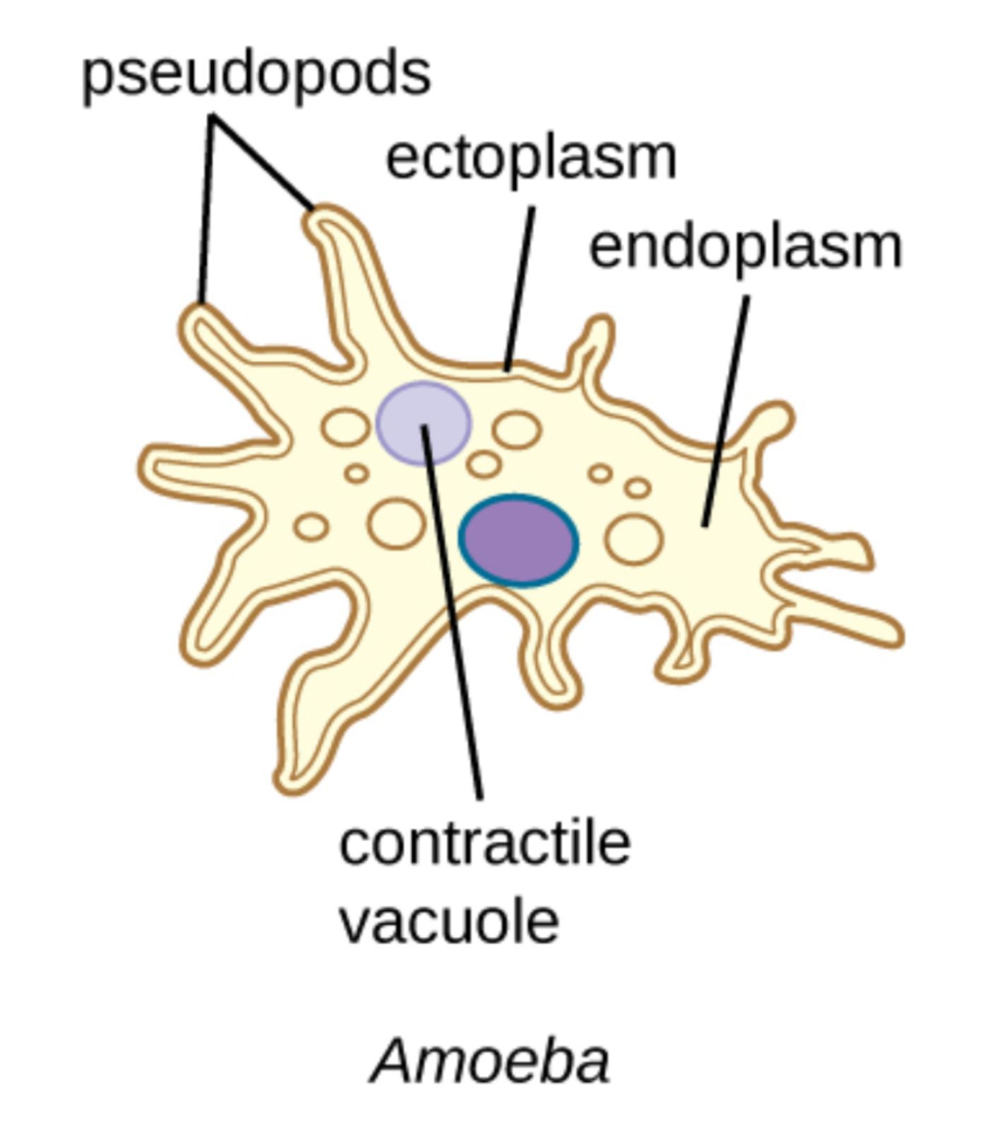
pseudopods: These are temporary, finger-like extensions of the cell membrane and cytoplasm used primarily for locomotion and the capture of food. They form as the cytoplasm flows in a specific direction, allowing the organism to essentially crawl along surfaces.
ectoplasm: This refers to the clear, non-granular outer layer of the cytoplasm located just beneath the cell membrane. It is more viscous and gel-like than the inner layer, providing structural support and playing a key role in the formation of pseudopodia.
endoplasm: This is the inner, more fluid, and granular portion of the cytoplasm which contains the nucleus, vacuoles, and other essential organelles. The constant streaming of this layer is what drives the internal pressure needed for the cell to change shape.
contractile vacuole: This specialized organelle acts as an osmoregulatory pump by collecting and periodically expelling excess water that enters the cell through osmosis. Without this mechanism, the freshwater-dwelling amoeba would eventually swell and burst due to internal pressure.
The Mechanics of Amoeboid Movement
One of the most fascinating aspects of this organism is its unique method of locomotion, often referred to as amoeboid movement. This process is driven by the internal transition of the cytoplasm between two states: the gel-like ectoplasm and the more fluid endoplasm. As the amoeba prepares to move, the endoplasm flows toward the leading edge of the cell, where it is converted into ectoplasm, expanding the pseudopod. Simultaneously, at the tail end of the cell, the ectoplasm is converted back into endoplasm to continue the forward flow. This continuous cycle of sol-gel transformation is powered by the same proteins found in human muscle cells—actin and myosin—demonstrating a highly conserved evolutionary mechanism for movement.
This movement is not merely random; it is often a directed response to environmental stimuli, such as chemical gradients or light. This behavior, known as chemotaxis, allows the amoeba to seek out nutrient-rich areas or avoid harmful substances. The flexibility afforded by the absence of a rigid cell wall makes the amoeba one of the most versatile navigators of the microscopic world, capable of squeezing through incredibly tight spaces in soil or tissue to reach its objectives.
Feeding and Digestion via Phagocytosis
Amoebae are heterotrophic organisms that sustain themselves by consuming bacteria, algae, and other smaller protozoa. The process by which they ingest food is known as phagocytosis, which literally translates to “cell eating.” When an amoeba detects a food particle, it extends its pseudopods to surround the prey. Eventually, the tips of the pseudopods meet and fuse, trapping the food along with a small amount of water within a membrane-bound sac called a food vacuole.
Once the food vacuole is internalized, digestive enzymes are secreted into the sac to break down the organic matter into simple molecules like glucose and amino acids. These nutrients then diffuse out of the vacuole into the surrounding cytoplasm to be used for energy and growth. Any indigestible remains are eventually moved toward the cell membrane, where the vacuole fuses with the exterior to expel the waste through exocytosis. This efficient intracellular digestive system allows a single cell to process a wide variety of food sources without the need for complex organ systems.
Internal Structure and Cytoplasmic Organization
Beneath the seemingly simple exterior of the amoeba lies a highly organized internal environment. The division between the ectoplasm and endoplasm is critical for both the cell’s integrity and its dynamic functions. The endoplasm acts as the metabolic hub, housing the nucleus—which contains the organism’s genetic material and coordinates growth and reproduction—as well as various mitochondria that produce the energy (ATP) required for cytoplasmic streaming. The granular appearance of the endoplasm is due to the presence of these organelles along with various crystals and storage granules.
The cell membrane, or plasmalemma, is thin and elastic, yet robust enough to protect the internal contents. It is semi-permeable, allowing for the exchange of gases like oxygen and carbon dioxide via simple diffusion. This direct interaction with the environment is essential for a unicellular organism that lacks specialized respiratory or circulatory systems. The constant rearrangement of the cytoskeleton within these layers allows the amoeba to maintain its physiological functions even while its outward form is in a state of perpetual change.
The Critical Role of Osmoregulation
Because most amoebae live in freshwater environments where the concentration of solutes is much higher inside the cell than outside, water constantly enters the organism through osmosis. To prevent the cell from rupturing, the amoeba employs osmoregulation through its contractile vacuole. This organelle slowly fills with excess water from the cytoplasm, expanding in size until it reaches a threshold. It then migrates to the edge of the cell, fuses with the plasma membrane, and forcefully contracts to expel the fluid.
The frequency of this contraction depends on the external environment; if an amoeba is placed in a more saline solution, the rate of water entry decreases, and the contractile vacuole pulses less often. This homeostatic mechanism is a primary indicator of cellular life and health. If the vacuole fails to function, the cell will rapidly experience osmotic shock. Studying this process helps researchers understand how cells manage ion balances and fluid pressure, which are vital concepts in human renal and cardiovascular medicine.
Reproduction Through Binary Fission
The primary method of reproduction for the amoeba is an asexual process called binary fission. This begins with the replication of the nucleus through mitosis, followed by the elongation of the entire cell. A constriction eventually forms in the middle of the cytoplasm, which deepens until the cell pinches into two separate, genetically identical daughter cells. This allows for rapid population growth when environmental conditions are favorable, such as an abundance of food and optimal temperatures.
Under less favorable conditions, some species of amoeba can undergo a process called encystment. The cell rounds up, secretes a thick, protective wall, and enters a dormant state. This cyst allows the organism to survive periods of drought or extreme temperature. When favorable conditions return, the cell emerges from the cyst and resumes its active life. This resilience is a key factor in the widespread geographical distribution of amoeboid species across the planet.
Clinical Significance and Pathogenic Amoebae
While many amoebae are harmless environmental scavengers, certain species are highly pathogenic to humans. The most notable is Entamoeba histolytica, which causes amoebic dysentery. This parasite invades the lining of the large intestine, leading to severe abdominal pain, diarrhea, and potentially life-threatening liver abscesses if it enters the bloodstream. Another dangerous relative is Naegleria fowleri, often called the “brain-eating amoeba,” which can cause primary amebic meningoencephalitis when contaminated water enters the nasal passages.
Understanding the fundamental anatomy and biological functions of the common amoeba provides the necessary groundwork for medical professionals to diagnose and treat these parasitic infections. Anti-parasitic medications often target the unique metabolic pathways or structural proteins of these organisms to eradicate them from the host. Furthermore, because amoebae utilize many of the same signaling and motility mechanisms as human white blood cells, they continue to be invaluable subjects in immunology and cancer research, where cellular invasion and movement are key areas of focus.
Summary of Key Biological Concepts
- Movement is achieved through pseudopodia and cytoplasmic streaming (sol-gel transition).
- Nutrition is managed via phagocytosis and the formation of food vacuoles.
- Homeostasis is maintained by the contractile vacuole’s active osmoregulation.
- Reproduction occurs primarily through asexual binary fission, with encystment as a survival tactic.
- Medical relevance stems from both their use as model cells and their potential as human pathogens.
The study of amoeboid life continues to reveal the hidden complexities of nature. From the coordinated flow of its internal fluids to its sophisticated hunting behaviors, the amoeba proves that even without a brain or complex organs, a single cell can exhibit highly adaptive and successful life strategies.
keywords: amoeba, pseudopods, ectoplasm, endoplasm, contractile vacuole, phagocytosis, binary fission, protozoa, osmoregulation, microbiology

