In the vast and intricate domain of microbiology, protozoa represent a diverse group of single-celled organisms that exhibit a complexity comparable to that of multicellular creatures. As protozoa are classified as eukaryotic cells, they possess membrane-bound organelles and a well-defined nucleus, allowing them to perform all the necessary functions of life within a single cellular boundary. These organisms are primarily distinguished and classified based on their unique modes of locomotion, which are dictated by specialized anatomical structures. From the slow, gliding movement of the amoeba to the rapid, rhythmic beating of a paramecium’s cilia, these microorganisms demonstrate evolutionary adaptations that allow them to thrive in diverse aquatic and moist terrestrial environments. Understanding their morphology is not merely a biological exercise; it provides essential insights into the mechanisms of cellular biology, osmoregulation, and the pathology of various human diseases caused by parasitic relatives of these common models.
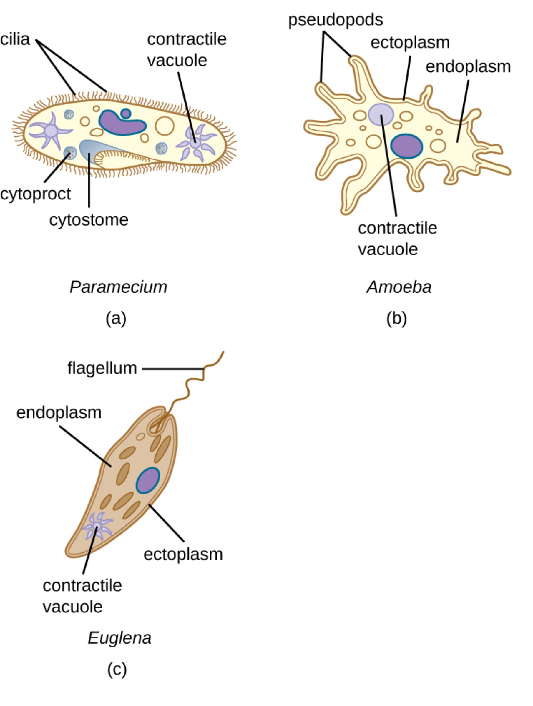
cilia: These are numerous, short, hair-like projections extending from the surface of the Paramecium that move in a coordinated wave-like pattern. They are primarily used for locomotion and to sweep food particles into the organism’s oral groove.
contractile vacuole: This specialized organelle acts as a pump to expel excess water that enters the cell through osmosis in freshwater environments. It is critical for maintaining the cell’s internal pressure and preventing it from bursting.
cytoproct: Also referred to as the anal pore, this structure serves as the exit point for indigestible waste materials. It ensures that the cellular digestive process maintains a consistent one-way flow of nutrients and waste.
cytostome: This is the specialized “cell mouth” found at the base of the oral groove in Paramecium. It facilitates the ingestion of food particles, which are then packaged into food vacuoles for internal digestion.
pseudopods: These are temporary, finger-like extensions of the Amoeba’s cell membrane and cytoplasm. They are utilized for “amoeboid movement” and for surrounding and engulfing prey through the process of phagocytosis.
ectoplasm: This is the thin, transparent, and relatively rigid outer layer of the cytoplasm located just beneath the plasma membrane. It provides structural support to the cell and plays a pivotal role in the initiation of pseudopod formation.
endoplasm: The granular and fluid-like inner portion of the cytoplasm where most of the cell’s metabolic activities occur. Its shifting internal pressure and movement drive the cytoplasmic streaming necessary for locomotion in Amoebae.
flagellum: A long, whip-like appendage that projects from the anterior end of the Euglena. By rotating or lashing this structure, the organism can propel itself forward through liquid media with significant speed.
The Evolutionary Success of Protozoan Locomotion
The classification of protozoa historically relies heavily on their locomotor organelles. The three primary modes of movement observed in the provided image—ciliary, amoeboid, and flagellar—represent sophisticated solutions to the challenges of moving through fluid environments at a microscopic scale. Paramecium, representing the Ciliophora phylum, uses thousands of cilia that are precisely synchronized through an internal network of fibers. This allows for rapid swimming and precise maneuvering, which is essential for an organism that actively hunts for bacteria. Amoebae, belonging to the Sarcodina group, utilize a more fluid approach through the formation of pseudopodia. This involves the rapid transformation of cytoplasm from a fluid “sol” state to a more rigid “gel” state, allowing the cell to literally flow across surfaces. Euglena, a member of the Mastigophora group, employs a flagellum. This structure is fundamentally different in its mechanics from cilia, providing a powerful pull that drags the organism toward light sources or nutrients.
Beyond movement, these locomotor structures often serve secondary functions. In Paramecium, the cilia surrounding the oral groove create a vortex that traps food particles. In Amoebae, pseudopods are essential for phagocytosis, where the cell membrane wraps around a food item to form an internal digestive vesicle. These adaptations highlight the efficiency of protozoa, where a single structure can handle movement, sensory perception, and nutrient acquisition simultaneously.
Osmoregulation and the Contractile Vacuole
One of the most significant physiological challenges faced by freshwater protozoa is the constant influx of water due to osmosis. Because the concentration of solutes inside the cell is higher than in the surrounding freshwater, water naturally diffuses across the semi-permeable cell membrane. Without a mechanism to counteract this, the cell would expand and eventually lyse. The osmoregulation process is managed by the contractile vacuole, an organelle visible in all three models: Paramecium, Amoeba, and Euglena. The vacuole functions through a cycle of diastole (filling) and systole (contracting). In Paramecium, the contractile vacuole is often surrounded by radiating canals that collect water from the cytoplasm and deliver it to the central reservoir. Once full, the vacuole fuses with the plasma membrane and forcefully ejects the water out of the cell. The rate of contraction is directly proportional to the salinity of the environment; in higher salinity, the vacuole pulses less frequently, demonstrating a highly responsive homeostatic mechanism.
Amoeba: The Dynamics of Cytoplasmic Streaming
The Amoeba is often studied as the simplest model of a eukaryotic cell, yet its internal dynamics are incredibly complex. The division of the cytoplasm into ectoplasm and endoplasm is not static. During movement, the fluid endoplasm flows toward the leading edge of a pseudopod. As it reaches the tip, it spreads out and thickens into the more rigid ectoplasm. Simultaneously, at the tail end of the cell, the ectoplasm liquefies into endoplasm to continue the flow. This “fountain zone” effect is powered by actin and myosin proteins, the same proteins responsible for muscle contraction in humans. This highlights a fundamental biological principle: the mechanical tools used by complex animals were first perfected in single-celled ancestors. Clinically, understanding the amoeboid movement is vital because many human immune cells, such as neutrophils and macrophages, use identical mechanisms to hunt and destroy pathogens within our tissues.
Paramecium: Complex Anatomy and Genetic Diversity
Paramecium species are arguably the most anatomically complex protozoa. Unlike the amorphous Amoeba, the Paramecium has a fixed, slipper-like shape maintained by a tough but flexible outer covering called the pellicle. This pellicle contains alveoli and the bases of the cilia (kinetosomes). Internally, Paramecium is unique because it possesses nuclear dualism. It has a large macronucleus that controls daily metabolic functions and a smaller micronucleus that is strictly reserved for reproduction. During the process of conjugation, two paramecia align and exchange genetic material via their micronuclei. This process does not increase the number of individuals but ensures genetic recombination, which is essential for surviving changing environmental pressures. The presence of specialized regions like the cytostome (mouth) and cytoproct (anus) shows a high degree of cellular differentiation, mirroring the organ systems of higher organisms.
Euglena: The Mixotrophic Hybrid
Euglena represents a fascinating evolutionary bridge between plants and animals. They are known as mixotrophs because they can function as both autotrophs and heterotrophs. In the presence of sunlight, the numerous chloroplasts within the endoplasm perform photosynthesis to produce energy. However, if light is absent, the Euglena can survive by absorbing organic nutrients from the water through its cell membrane. To aid in its photosynthetic lifestyle, the Euglena possesses a specialized light-sensing organelle near the base of the flagellum called the stigma, or eyespot. The stigma shields a light-sensitive swelling on the flagellum, allowing the organism to detect the direction of light and swim toward it (positive phototaxis). This dual lifestyle makes Euglena an exceptionally resilient organism, capable of thriving in environments where other specialized microorganisms might fail.
Medical Significance of Protozoan Structures
While the models shown in the image are generally non-pathogenic, they are closely related to significant human pathogens. For instance, the mechanisms of pseudopod formation in the common Amoeba are identical to those used by Entamoeba histolytica, the causative agent of amoebic dysentery, which invades the intestinal wall. Similarly, the flagellar mechanics of Euglena provide a blueprint for understanding Trypanosoma, the parasite responsible for African Sleeping Sickness, which uses its flagellum to navigate the human bloodstream. Many anti-parasitic drugs target the very structures seen in this image, such as disrupting the microtubules in cilia/flagella or interfering with the osmoregulatory functions of the contractile vacuole. By studying these non-pathogenic models, researchers can develop strategies to combat infectious diseases without the risks associated with handling highly virulent strains.
Summary of Protozoan Comparison
- Locomotion: Cilia (Paramecium), Pseudopods (Amoeba), Flagella (Euglena).
- Feeding: Oral groove/Cytostome (Paramecium), Phagocytosis (Amoeba), Photosynthesis/Absorption (Euglena).
- Excretion: Cytoproct (Paramecium), Diffusion/Exocytosis (Amoeba and Euglena).
- Shape: Fixed slipper shape (Paramecium), Constantly changing (Amoeba), Spindle-shaped/Flexible (Euglena).
- Environment Control: All use contractile vacuoles for active water expulsion.
In conclusion, the microscopic world of protozoa is a testament to the versatility of the eukaryotic cell. Whether through the coordinated rhythm of cilia, the fluid grace of pseudopodia, or the powerful pull of a flagellum, these organisms have mastered the art of survival. Their structural features—from the protective pellicle to the complex contractile vacuole—serve as the foundation for our understanding of cellular physiology and the evolutionary history of life on Earth.

