Bacillus cereus is a widespread, spore-forming Gram-positive bacterium commonly encountered in soil, food, and the environment, known for causing foodborne illnesses and occasional opportunistic infections. Its identification in the laboratory relies heavily on microscopic examination after Gram staining, where it displays characteristic large rod-shaped cells often arranged in chains. The image provides a detailed microscopic view of this organism under oil immersion, highlighting the typical morphology that distinguishes it from other bacilli and aiding students, microbiologists, and clinicians in accurate recognition.
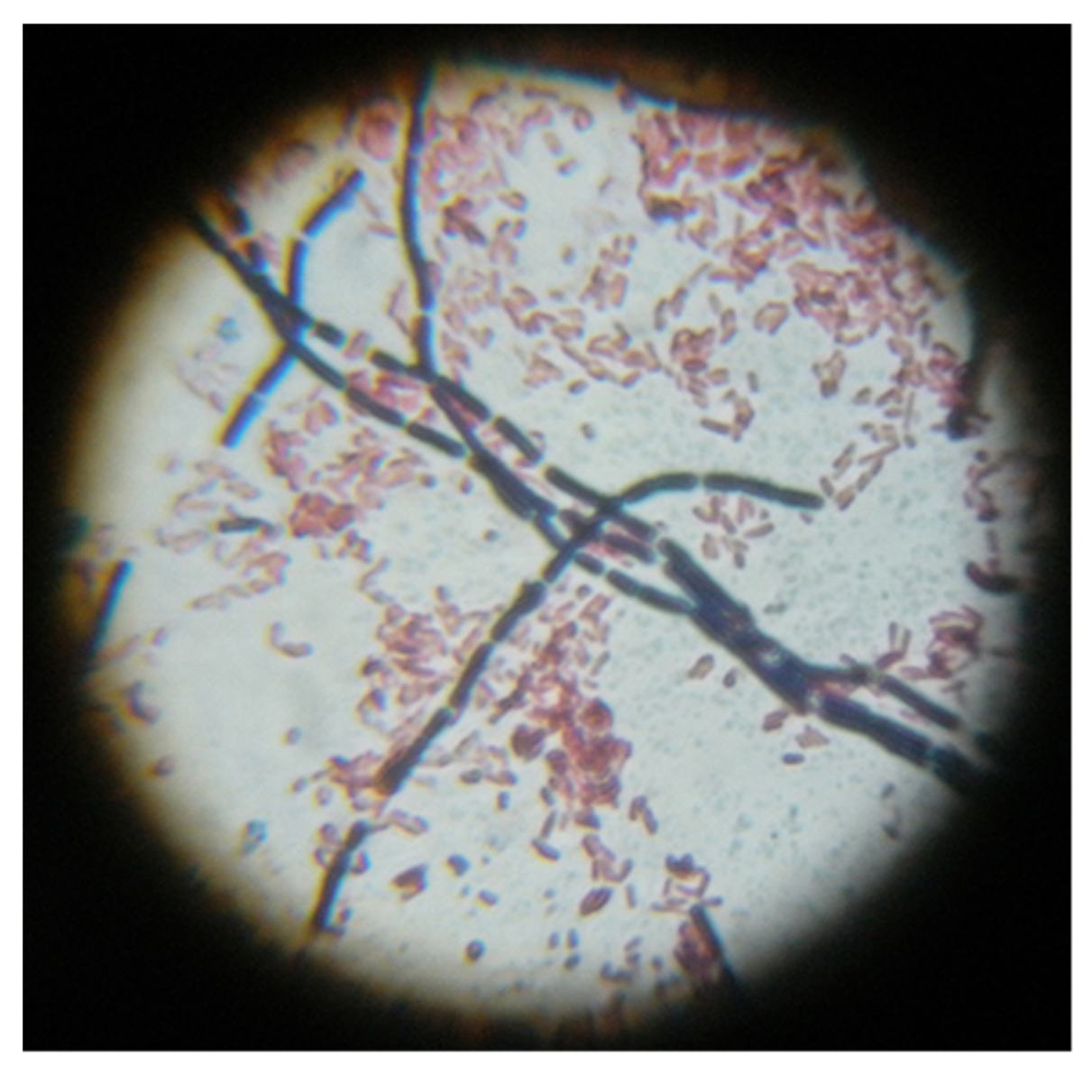
Gram-positive rods appear as large, violet-stained bacilli with square or slightly rounded ends in the image. These cells retain the crystal violet-iodine complex due to their thick peptidoglycan layer, resulting in the deep purple coloration visible against the lighter background. Bacillus cereus rods measure approximately 1-1.5 μm in width by 3-5 μm or longer in length and frequently form chains, giving a bamboo-like appearance with clear junctions between individual cells.
Chains of bacilli are prominently displayed as elongated, interconnected violet structures crossing the field of view. This arrangement occurs because daughter cells remain attached after division, especially in fresh cultures. The image captures both long serpentine chains and shorter segments, a hallmark feature that helps differentiate B. cereus from non-chain-forming Gram-positive rods during preliminary microscopic evaluation.
Pink background cells represent smaller Gram-negative bacteria or debris that have taken up the safranin counterstain. These pink elements provide contrast, emphasizing the larger size and stronger staining of the Gram-positive Bacillus cereus rods. In mixed specimens, such as the one shown, this differential staining underscores the importance of Gram reaction in guiding further identification and ruling out contaminants.
Morphological Features of Bacillus cereus Under Microscopy
When viewed after standard Gram staining, Bacillus cereus presents as large, straight or slightly curved rods with square ends. The cells are typically arranged singly, in pairs, or in short to long chains, as clearly illustrated in the image. This morphology is consistent across most culture conditions, though older cultures may occasionally appear Gram-variable due to cell wall changes during sporulation or autolysis.
The thick peptidoglycan layer characteristic of Gram-positive bacteria allows strong retention of the primary stain, producing the intense violet hue seen throughout the micrograph. Endospores, when present, are ellipsoidal and positioned centrally or subterminally without significantly swelling the sporangium; however, they may not always be visible in routine Gram stains without specialized spore staining techniques.
- Rods measure roughly 1 × 3-5 μm, making them noticeably larger than many other common bacilli.
- Chains often resemble bamboo stalks with distinct septa between cells.
- In clinical specimens or tissue sections, the organism can appear more filamentous or elongated.
Laboratory Identification and Cultural Characteristics
Gram staining serves as the initial step in identifying Bacillus cereus from clinical or food samples. The image exemplifies the classic appearance that prompts further testing on selective or differential media such as mannitol egg yolk polymyxin (MYP) agar or sheep blood agar. On blood agar, colonies are large, flat, irregular, and often beta-hemolytic, with a ground-glass texture.
Biochemical confirmation includes positive catalase, lecithinase production (opaque zones on egg yolk agar), motility, and Voges-Proskauer reaction. Molecular methods targeting toxin genes or 16S rRNA sequencing provide definitive identification when needed. The microscopic features shown help distinguish B. cereus from closely related species like B. anthracis, which produces non-hemolytic colonies and lacks motility.
- Growth occurs readily on nutrient agar at 30-37°C under aerobic or facultative anaerobic conditions.
- Endospore formation allows survival of heat, desiccation, and disinfectants, complicating food safety efforts.
- Penicillin resistance due to beta-lactamase production is a useful differentiating trait.
Pathogenesis and Clinical Significance of Bacillus cereus
Bacillus cereus is best known for two distinct food poisoning syndromes. The emetic syndrome results from ingestion of preformed heat-stable cereulide toxin, typically in rice or pasta dishes, causing rapid-onset nausea and vomiting within hours. The diarrheal syndrome involves production of heat-labile enterotoxins in the intestine after consumption of contaminated food, leading to watery diarrhea and abdominal cramps after a longer incubation period.
In addition to gastroenteritis, the organism can cause severe opportunistic infections, including ocular infections such as endophthalmitis following trauma, as well as bacteremia, pneumonia, and wound infections in immunocompromised patients. Its ability to form biofilms and spores contributes to persistence in hospital environments and resistance to some sterilization methods.
Virulence factors include multiple enterotoxins, hemolysins, phospholipases, and proteases that damage host tissues. The robust cell wall and spore-forming capability enable the bacterium to withstand harsh conditions, explaining its frequent isolation from a variety of food products and environmental sources.
- Emetic toxin is a small cyclic peptide extremely resistant to heat, acid, and proteolysis.
- Diarrheal toxins are proteins sensitive to heat and produced in vivo.
- Invasive disease is more common in patients with indwelling devices or immunosuppression.
Prevention, Treatment, and Public Health Implications
Prevention of Bacillus cereus foodborne illness centers on proper food handling practices: rapid cooling of cooked foods, thorough reheating, and avoidance of prolonged room-temperature storage of potentially contaminated items. In healthcare settings, strict infection control and equipment sterilization reduce the risk of nosocomial infections.
Most cases of food poisoning are self-limited and managed with supportive care, including hydration. Severe or invasive infections require antibiotic therapy, often with vancomycin, clindamycin, or fluoroquinolones, selected according to susceptibility testing. Early diagnosis, supported by recognition of the Gram stain morphology shown in the image, is critical for optimal outcomes in serious cases.
Public health surveillance monitors outbreaks linked to contaminated food products, particularly in institutional settings like schools or restaurants. Education on safe food preparation remains the most effective strategy to minimize the impact of this environmentally persistent pathogen.
The microscopic image serves as an essential educational tool, bridging the gap between theoretical knowledge and practical laboratory skills. By familiarizing viewers with the distinctive chains of large Gram-positive rods, it enhances the ability of laboratory personnel to rapidly identify potential B. cereus isolates and initiate appropriate confirmatory testing.
In summary, understanding the Gram stain appearance of Bacillus cereus is fundamental to clinical microbiology. The clear visualization of rod-shaped cells in chains provides immediate clues that guide further investigation, ultimately contributing to accurate diagnosis, effective treatment, and prevention of associated diseases.

