Bacillus cereus stands as a ubiquitous, spore-forming Gram-positive bacterium frequently implicated in foodborne illnesses and opportunistic infections. Commonly found in soil, vegetation, and various food products, this hardy organism demonstrates distinctive microscopic and cultural characteristics that aid in its laboratory identification. The provided image beautifully illustrates both the Gram-stained cellular morphology in a mixed preparation and the typical colony appearance on sheep blood agar, offering valuable insights into the identification and behavior of B. cereus in clinical and food microbiology settings.
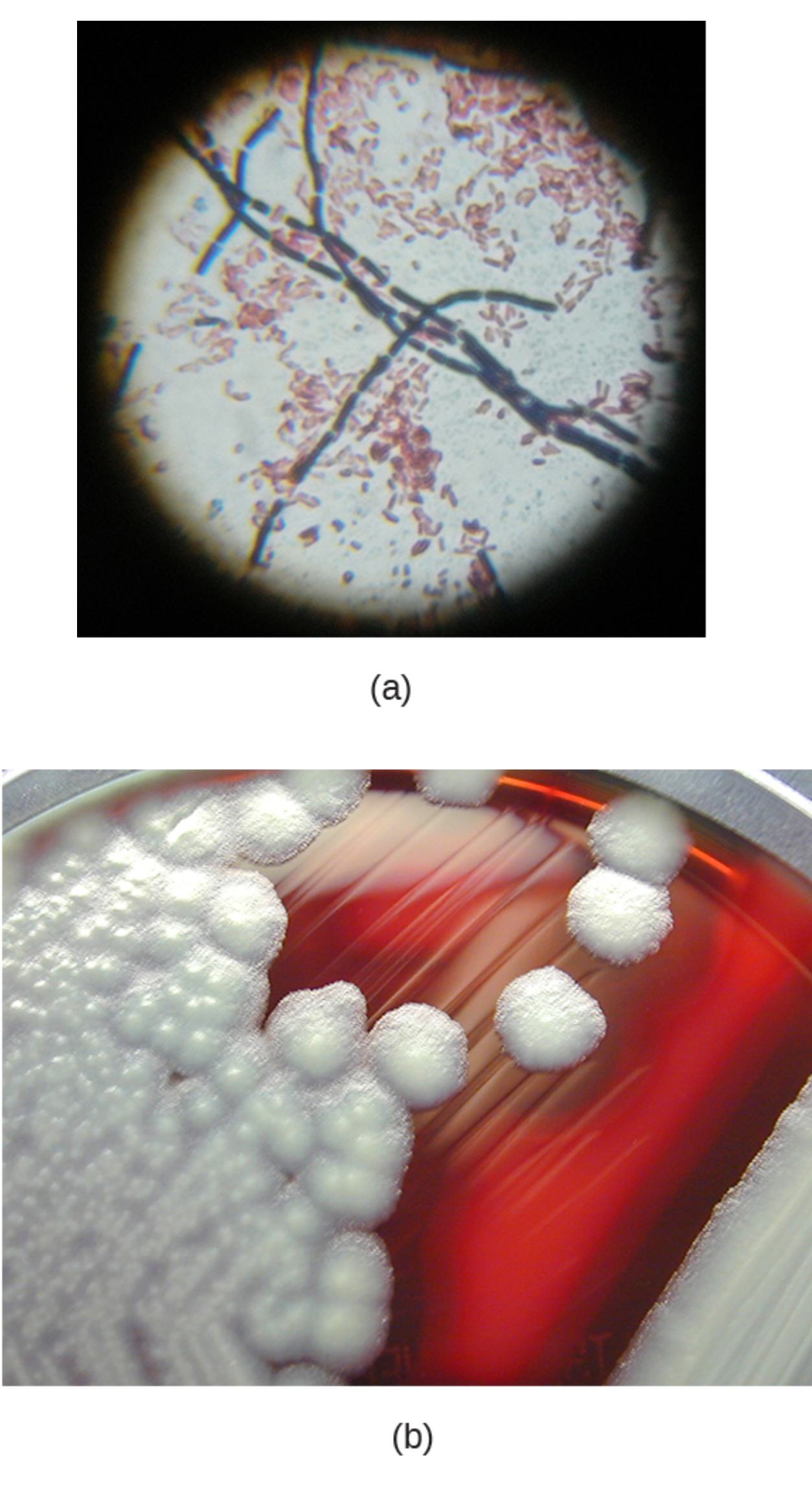
Gram-positive bacteria in the top panel (a) appear as violet rod-shaped cells arranged singly or in chains, characteristic of Bacillus cereus. These large bacilli retain the crystal violet stain due to their thick peptidoglycan layer in the cell wall. In the mixed specimen, they contrast sharply with smaller pink-staining organisms, highlighting the differential staining property that separates Gram-positive from Gram-negative bacteria.
Escherichia coli are the small, pink rod-shaped cells visible in the Gram-stained preparation (a). As a classic Gram-negative bacterium, E. coli loses the primary crystal violet stain during decolorization and takes up the safranin counterstain. This mixture serves as an excellent teaching tool to demonstrate side-by-side comparison of Gram reaction, cell size, and morphology between the two species.
White colonies in the bottom panel (b) represent the growth of B. cereus on sheep blood agar. These colonies typically appear large, opaque, and somewhat irregular or feathery with a rough, matted texture. Their white to grayish appearance against the red agar background is a common feature observed after 24 hours of incubation at 37°C, often accompanied by surrounding hemolytic activity.
Sheep blood agar provides a nutrient-rich medium that supports robust growth of B. cereus while allowing observation of hemolytic patterns. The red coloration comes from incorporated sheep erythrocytes, which the bacterium can lyse through production of hemolysins. In the image, the medium displays clear differentiation between the dense white bacterial growth on the left and individual well-defined colonies on the right.
Microbiological Characteristics of Bacillus cereus
Bacillus cereus is a facultative anaerobe and spore-former belonging to the Bacillus cereus group. It measures approximately 1 × 3-5 μm with square or slightly rounded ends and often appears in chains. On Gram stain, fresh cultures reliably show Gram-positive rods, though older cultures may appear Gram-variable. The top panel of the image effectively demonstrates this morphology alongside Escherichia coli for contrast.
Colony morphology on blood agar is particularly distinctive. Colonies are large (2-5 mm or more), flat to slightly raised, with irregular edges and a ground-glass or frosted appearance. They frequently exhibit beta-hemolysis, producing clear zones around colonies due to toxin-mediated red blood cell destruction. The bottom panel (b) captures the typical white, opaque colonies that spread or swarm under certain conditions.
- Endospores are central to subterminal and do not distend the cell significantly.
- The organism is catalase positive and motile via peritrichous flagella.
- Growth occurs over a wide temperature range, with optimal activity around 30-37°C.
Pathogenesis and Food Poisoning Syndromes
Bacillus cereus produces multiple toxins responsible for two main forms of foodborne illness: the emetic syndrome and the diarrheal syndrome. The emetic type, caused by the preformed heat-stable cereulide toxin, leads to rapid-onset nausea and vomiting within 0.5 to 6 hours after ingestion, often linked to starchy foods like rice or pasta left at room temperature. The diarrheal form involves heat-labile enterotoxins (such as hemolysin BL and non-hemolytic enterotoxin) produced in the intestine, resulting in abdominal cramps and watery diarrhea 8-16 hours post-exposure.
Beyond food poisoning, B. cereus can cause serious opportunistic infections, particularly in immunocompromised individuals. These include ocular infections (endophthalmitis), bacteremia, pneumonia, and wound infections. Its ability to form biofilms and spores contributes to persistence in hospital environments and resistance to certain disinfectants.
- Emetic toxin is a small cyclic peptide highly resistant to heat, acid, and enzymes.
- Diarrheal enterotoxins are proteins destroyed by heating above 56°C for 30 minutes.
- Other virulence factors include phospholipases, proteases, and hemolysins.
Laboratory Identification and Differentiation
Identification of Bacillus cereus begins with Gram staining and culture on selective or differential media. The image’s top panel showcases the classic Gram-positive rods in contrast to Gram-negative Escherichia coli, reinforcing the importance of staining in preliminary identification. On sheep blood agar, the large beta-hemolytic colonies help distinguish it from non-hemolytic species like Bacillus anthracis.
Further confirmation involves biochemical tests such as positive lecithinase activity on egg yolk agar (producing an opaque zone), positive Voges-Proskauer reaction, and motility. Commercial systems or molecular methods targeting toxin genes provide rapid and specific identification. The white colonies shown in the bottom panel are typical but must be correlated with hemolysis and other traits to avoid confusion with other Bacillus species.
- Selective media like mannitol egg yolk polymyxin (MYP) agar yield pink colonies with lecithinase halos.
- B. cereus is penicillin resistant due to beta-lactamase production, unlike B. anthracis.
- Spore staining reveals central or subterminal endospores that survive harsh conditions.
Clinical Management and Prevention
Most cases of B. cereus food poisoning are self-limiting and require only supportive care, including hydration. Severe or invasive infections necessitate antibiotics such as vancomycin, clindamycin, or fluoroquinolones, guided by susceptibility testing. Early recognition is critical in ocular or systemic cases to prevent permanent damage.
Prevention focuses on proper food handling: rapid cooling of cooked foods, reheating to at least 74°C, and avoiding prolonged storage at room temperature. In healthcare settings, strict infection control measures reduce the risk of nosocomial transmission from contaminated equipment or solutions.
The dual panels in the image serve as powerful educational resources, linking cellular morphology observed under the microscope with macroscopic colony features on culture media. Understanding these characteristics enables microbiologists and clinicians to quickly recognize and address potential B. cereus contamination or infection.
Overall, Bacillus cereus exemplifies the dual nature of many environmental bacteria—harmless or beneficial in some contexts yet capable of causing significant disease when conditions favor toxin production or host vulnerability. Continued vigilance in food safety and laboratory diagnostics remains essential to mitigate its impact on public health.

