Jugular Venous Distension (JVD) is a critical clinical sign often observed in patients with significant cardiovascular compromise, serving as a window into the hemodynamics of the right side of the heart. The image provided illustrates a classic presentation of elevated venous pressure in the neck of an elderly male patient, acting as a vital diagnostic clue for healthcare providers assessing fluid status and cardiac function. By observing the distinct bulging of the neck veins, clinicians can estimate the central venous pressure without invasive procedures, aiding in the diagnosis of conditions such as heart failure.
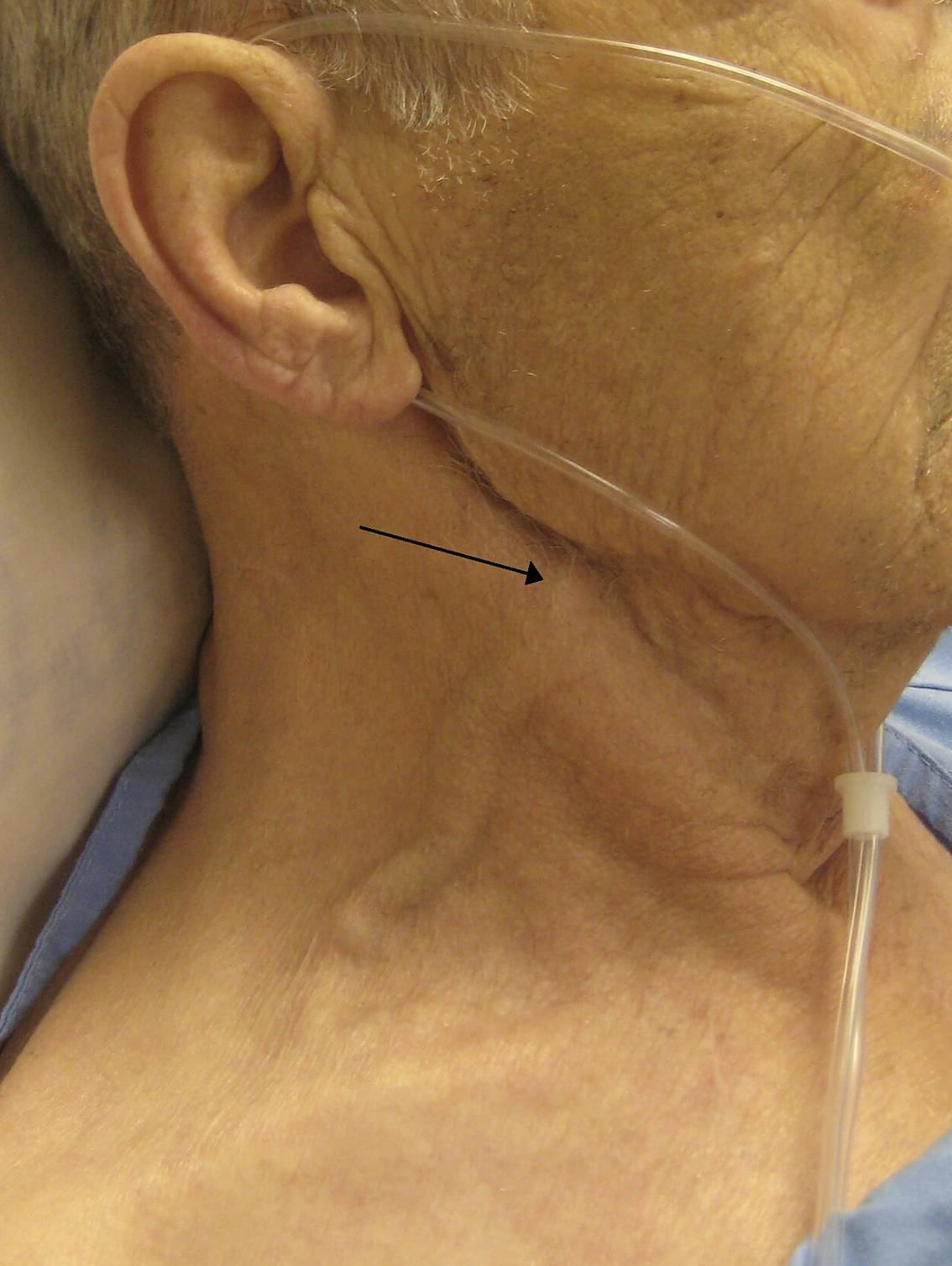
Arrow pointing to vein: The black arrow in the image specifically highlights the external jugular vein, which runs superficially across the sternocleidomastoid muscle. While this vein is visibly distended due to increased pressure backing up from the heart, it is important to note that clinical measurements of central venous pressure are typically calculated using the pulsations of the internal jugular vein, which is also visible in this patient’s neck but located deeper.
The Significance of Elevated Jugular Venous Pressure
The jugular veins act as a manometer for the right atrium of the heart, providing a direct visual representation of the pressure within the central venous system. In a healthy individual, the jugular veins are usually not visible when the patient is sitting upright or at a 45-degree angle. However, when the pressure in the right atrium rises, blood backs up into the superior vena cava and subsequently into the jugular veins, causing them to bulge or distend. This phenomenon is known as Jugular Venous Distension (JVD).
Physiologically, the absence of valves between the internal jugular vein and the right atrium allows for this direct transmission of pressure. Therefore, the height of the venous column in the neck correlates with the volume and pressure status of the right heart. When a clinician assesses a patient, they look for the double waveform pulsation of the internal jugular vein to distinguish it from the carotid artery, which has a single, palpable pulsation.
There are several pathological conditions that can lead to the elevation of Jugular Venous Pressure. While heart failure is the most common culprit, other conditions that obstruct blood flow into the heart or increase thoracic pressure can also cause this sign. Identifying JVD is often one of the first steps in a physical examination for patients presenting with shortness of breath or edema.
Common causes of elevated JVP include:
- Right-sided heart failure
- Tricuspid valve stenosis or regurgitation
- Cardiac tamponade (fluid accumulation around the heart)
- Constrictive pericarditis
- Pulmonary hypertension
- Superior vena cava obstruction
Congestive Heart Failure: Pathology and Presentation
The patient in the image is suffering from Congestive Heart Failure (CHF), a chronic condition where the heart muscle is unable to pump blood efficiently enough to meet the body’s needs for blood and oxygen. While CHF can affect the left side, the right side, or both, the visual sign of JVD is most directly associated with right-sided heart failure.
In left-sided heart failure, the left ventricle struggles to pump blood out to the body, causing fluid to back up into the lungs (pulmonary edema). As the pressure in the pulmonary circulation increases, the right ventricle must work harder to push blood into the lungs. Eventually, the right ventricle may weaken and fail, leading to right-sided heart failure. When the right ventricle cannot empty effectively, blood backs up into the right atrium and then into the systemic veins, resulting in the distended neck veins seen in the photograph.
Beyond the visible neck veins, patients with CHF often present with a constellation of symptoms related to fluid overload and poor perfusion. Systemic congestion can lead to peripheral edema (swelling in the legs and ankles), ascites (fluid in the abdomen), and hepatomegaly (liver enlargement). The diagnostic assessment often involves an echocardiogram to measure the ejection fraction and visualize valve function, alongside blood tests for biomarkers like B-type natriuretic peptide (BNP), which is elevated when the heart is under stress.
Distinguishing the External and Internal Jugular Veins
The image provides a valuable educational distinction between the external and internal jugular veins. The arrow marks the external jugular vein, which is more superficial and easier to visualize because it sits closer to the skin surface. However, the external jugular vein is prone to kinking and has valves that can make it a less reliable manometer for precise pressure measurement.
Conversely, the internal jugular vein runs deep to the sternocleidomastoid muscle and provides a straight path to the right atrium. While less visually distinct than the bulging external vein shown, the internal jugular vein’s pulsations are the gold standard for measuring central venous pressure at the bedside. In severe cases of CHF, as seen here, the pressure is so high that both venous systems become engorged, making the pathology unmistakable even to the naked eye.
In conclusion, the bulging vein depicted in the image is a hallmark sign of systemic venous congestion secondary to congestive heart failure. It serves as a powerful reminder of how external physical signs can reveal internal hemodynamic struggles. For medical professionals, recognizing and interpreting JVD is an essential skill that guides the management of diuretic therapy and the overall stabilization of heart failure patients.

