Total knee replacement, or total knee arthroplasty, is a definitive surgical solution for end-stage joint degeneration, resulting in a significant post-operative incision that requires careful management. This article explores the visual characteristics of a stapled surgical wound following knee replacement, the underlying pathology of osteoarthritis that necessitates this procedure, and the physiological stages of tissue healing.
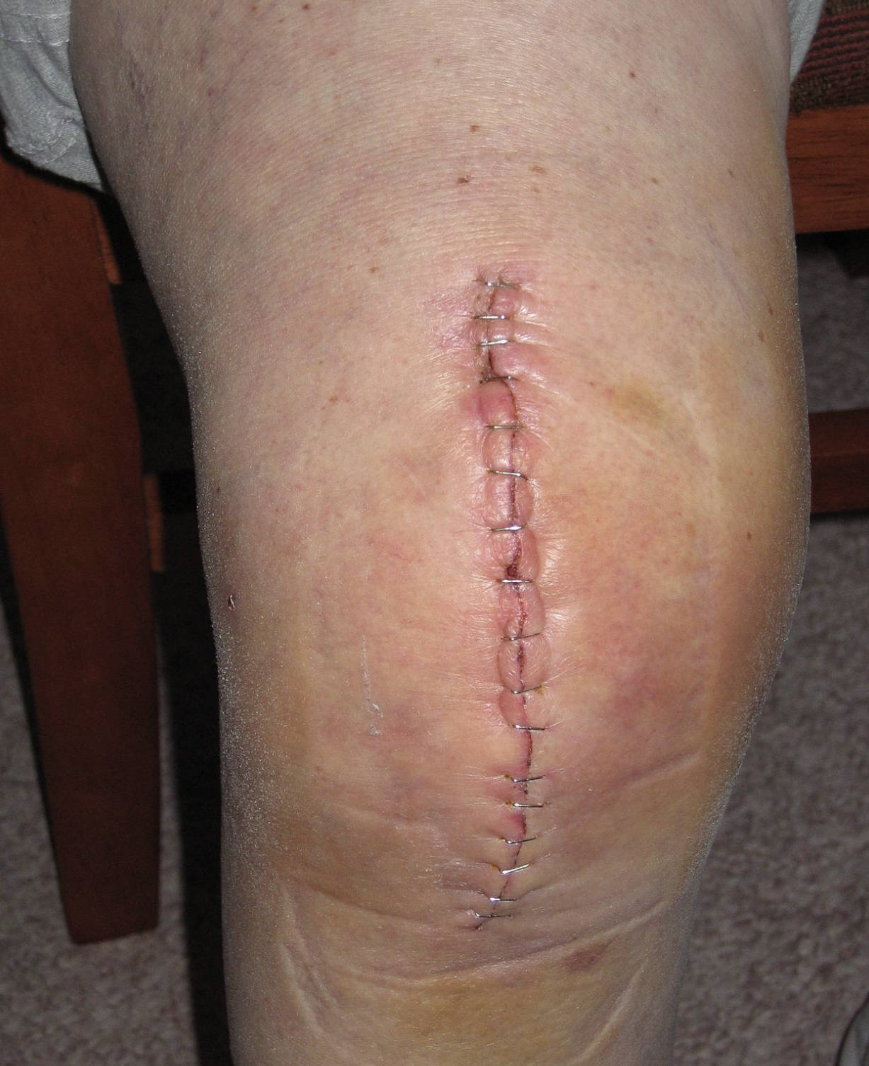
Surgical Staples: These are the metal fasteners visible along the length of the incision, used to hold the skin edges together. Staples are frequently chosen for orthopedic surgeries because they are quick to apply, reduce the risk of infection, and provide strong tension resistance for a joint that needs to bend.
Midline Incision: This label refers to the vertical cut running down the center of the knee, which is the standard surgical approach for a total knee replacement. The incision is typically 4 to 6 inches long, allowing the surgeon adequate access to resurface the distal femur and proximal tibia.
Peri-wound Erythema: This term describes the slight pink or red discoloration seen on the skin immediately surrounding the staples. This is a normal part of the inflammatory phase of healing, caused by increased blood flow to the area to deliver immune cells and nutrients for repair.
Post-Operative Wound Management in Orthopedics
The image displays a classic example of a primary intention wound closure following a total knee arthroplasty. In this procedure, the surgeon makes a vertical incision to access the knee joint, removes damaged cartilage and bone, and replaces them with artificial components. Once the internal structures are secured, the layers of tissue—the joint capsule, muscle, and subcutaneous fat—are stitched closed with absorbable sutures. The final layer, the epidermis, is often closed with stainless steel staples as seen here, which are typically removed 10 to 14 days post-surgery.
The appearance of the wound in the image suggests it is in the early stages of healing, likely within the first week after the operation. The skin edges are approximated (brought together) cleanly, which is essential for minimizing scarring and preventing pathogen entry. The knee is a high-tension area because the skin stretches tightly when the joint bends; therefore, the closure method must be robust enough to withstand early physical therapy while still allowing for adequate blood supply to the healing tissues.
Proper care of this surgical site is paramount to the success of the knee replacement. Patients are generally advised to keep the area clean and dry to prevent surgical site infections (SSIs). While some redness and swelling are normal, significant changes can indicate complications.
Key signs of proper healing versus potential complications include:
- Normal Healing: Gradual reduction in redness, minimal clear drainage, and decreasing tenderness.
- Infection Warning Signs: Spreading redness (cellulitis), thick yellow or green discharge (purulence), increasing pain, or fever.
- Dehiscence: The splitting open of the wound edges, which is a medical emergency.
- Staple embedding: If swelling is excessive, staples can dig into the skin, requiring medical assessment.
The Pathology Necessitating Surgery: Osteoarthritis
The primary indication for a total knee replacement is severe, debilitating osteoarthritis. This is a degenerative joint disease characterized by the progressive breakdown of articular cartilage. In a healthy knee, cartilage acts as a smooth, slippery cushion covering the ends of the femur (thigh bone) and tibia (shin bone), allowing for frictionless movement. Over time, due to age, trauma, or genetic factors, this cartilage wears away. Eventually, the protective layer disappears entirely, leading to a “bone-on-bone” state.
When the raw bone surfaces rub against each other, it triggers chronic inflammation, the formation of bone spurs (osteophytes), and severe pain. The physiological response involves the thickening of the joint capsule and the weakening of surrounding muscles due to disuse. Patients often experience stiffness, a reduced range of motion, and a grinding sensation known as crepitus. When conservative treatments like physical therapy, corticosteroid injections, and weight loss fail to provide relief, replacing the joint surfaces is the only viable option to restore quality of life.
Physiology of Surgical Incision Healing
The healing process shown in the image follows a predictable biological timeline. The first phase, hemostasis, occurs immediately during surgery, where platelets form a clot to stop bleeding. This is followed by the inflammatory phase, which is visible in the image as slight swelling and redness. During this time, white blood cells, particularly neutrophils and macrophages, migrate to the incision to destroy any bacteria and clear away cellular debris. This phase usually peaks within a few days of surgery.
Following inflammation, the proliferative phase begins. Fibroblasts enter the wound site and begin synthesizing collagen, the structural protein that knits the skin edges together. New blood vessels also form (angiogenesis) to supply oxygen to the regenerating tissue. Finally, the remodeling phase occurs, which can last for over a year. During this time, the collagen fibers reorganize to become stronger, and the red, raised scar gradually flattens and fades to a silvery-white line. The use of surgical staples facilitates the initial phases by keeping the skin edges in close contact, minimizing the distance the new tissue needs to bridge.
Conclusion
The stapled incision depicted represents the crucial final step of a Total Knee Arthroplasty, acting as the gateway to restored mobility. While the metal staples may look stark, they play a vital role in ensuring the skin heals securely over the new prosthetic joint. Understanding the anatomy of the closure and the physiology of the underlying healing process helps patients and caregivers recognize the difference between normal recovery and signs of complication, ensuring the best possible long-term outcome for the reconstructed knee.

