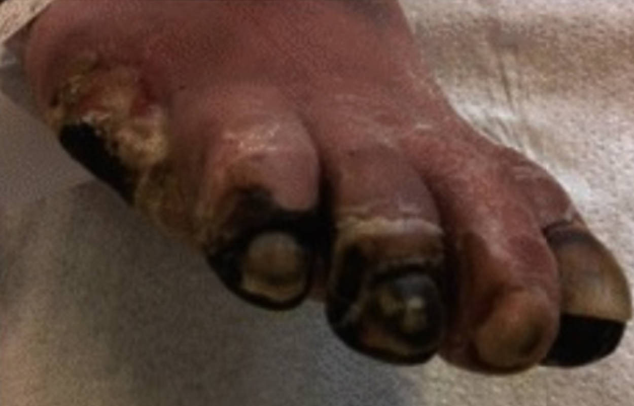
Peripheral Arterial Disease (PAD) is a progressive circulatory disorder characterized by the narrowing or blockage of the arteries supplying blood to the limbs, most commonly the legs and feet. When this condition reaches an advanced stage, known as critical limb ischemia, the complete cessation of blood flow results in tissue death. The image provided illustrates a severe manifestation of this disease, where peripheral arterial disease has led to necrosis (gangrene) of multiple toes, evidenced by the blackening and mummification of the tissue due to prolonged oxygen deprivation.
The Progression from Ischemia to Gangrene
The image depicts the devastating physical consequences of untreated or aggressive vascular disease. The toes appear black, shriveled, and hardened, a condition clinically referred to as dry gangrene. This occurs when the arterial blood supply is gradually cut off, preventing oxygen and nutrients from reaching the distal extremities. Without a blood supply, the cells undergo necrosis (death), and the tissue desiccates, or dries out. Unlike wet gangrene, which involves bacterial infection and swelling, dry gangrene is primarily an ischemic process where the tissue essentially mummifies.
This state is the culmination of a long-term pathological process. It typically begins with atherosclerosis, where fatty deposits accumulate in the arterial walls. Over years, this plaque hardens and narrows the arteries (stenosis). Initially, patients may experience pain while walking (claudication), but as the lumen of the artery becomes critically narrow or fully occluded, the blood flow is insufficient even at rest. This leads to rest pain, ulceration, and finally, the frank necrosis visible in the image.
Key clinical features often associated with the pathology shown above include:
- Color Change: The tissue transitions from pale or cyanotic (blue) to purple, and finally to black as hemoglobin breaks down and tissue dies.
- Demarcation: A distinct line often forms separating the healthy, viable tissue from the dead, necrotic tissue.
- Temperature Gradient: The affected foot or toes will feel noticeably cold to the touch due to the lack of perfusion.
- Sensory Loss: Patients often experience numbness or paresthesia, sometimes masking the pain of the dying tissue.
Pathophysiology of Peripheral Arterial Disease
The underlying cause of the necrosis shown is atherosclerosis, a systemic disease affecting the arterial tree. It begins with damage to the endothelium, the inner lining of the blood vessel, often caused by risk factors such as smoking, high blood pressure (hypertension), high cholesterol (hyperlipidemia), and diabetes mellitus. In response to this injury, the body attempts to repair the damage, but this leads to the accumulation of lipids, calcium, and inflammatory cells, forming a plaque.
As these plaques grow, they encroach upon the vessel lumen, increasing resistance to blood flow. In the lower extremities, the femoral, popliteal, and tibial arteries are frequently affected. When the diameter of the artery is reduced by more than 70%, the flow is significantly compromised. In the case of the image, the occlusion is likely located in the distal arteries (those in the calf or foot) or is a result of multi-level disease, meaning blockages exist in the thigh and the calf simultaneously. This multi-level obstruction makes it nearly impossible for the heart to pump blood all the way to the toes.
Diabetes mellitus significantly exacerbates this process. High blood sugar levels accelerate arterial stiffening (arteriosclerosis) and often affect the smaller vessels below the knee, making surgical reconstruction more difficult. Furthermore, diabetic patients frequently suffer from peripheral neuropathy (nerve damage), meaning they may not feel the initial pain of ischemia or minor trauma, allowing the condition to progress to gangrene before medical attention is sought.
Clinical Management and Treatment
The presence of necrotic tissue as seen in the image represents a medical urgency. The goals of treatment are threefold: to preserve as much of the limb as possible, to relieve pain, and to prevent the spread of necrosis or secondary infection. Diagnostic imaging, such as an angiogram or CT angiography, is performed to map the arterial anatomy and identify blockages. If feasible, revascularization is the priority. This involves restoring blood flow through endovascular procedures (angioplasty and stenting) or open surgical bypass grafting.
However, for the specific toes that have already turned black, the tissue is non-viable and cannot be saved. Once revascularization restores flow to the remaining healthy part of the foot, the dead tissue must be removed. This can occur through auto-amputation (where the body naturally sheds the dry, dead tissue) or, more commonly, surgical amputation. The level of amputation—whether it is just the toes, the forefoot, or higher—depends entirely on how far the blood flow can be restored. If revascularization fails or is not possible, major limb amputation (below or above the knee) may be necessary to prevent systemic infection and death.
Conclusion
The image of necrotic toes serves as a stark indicator of the severity of critical limb ischemia resulting from peripheral arterial disease. It highlights the mechanical failure of the circulatory system to sustain living tissue. While modern medicine offers advanced techniques to restore blood flow, the presence of gangrene signals that the disease is in its end stages. Early detection of risk factors, aggressive management of diabetes and hypertension, and smoking cessation remain the most effective tools in preventing such drastic outcomes.

