This transmission electron micrograph reveals the distinctive filamentous structure of the Ebolavirus, a highly pathogenic member of the Filoviridae family. By examining its physical morphology, medical professionals gain insight into how this deadly virus operates, identifying key features that contribute to its ability to cause severe hemorrhagic fever in humans and nonhuman primates.
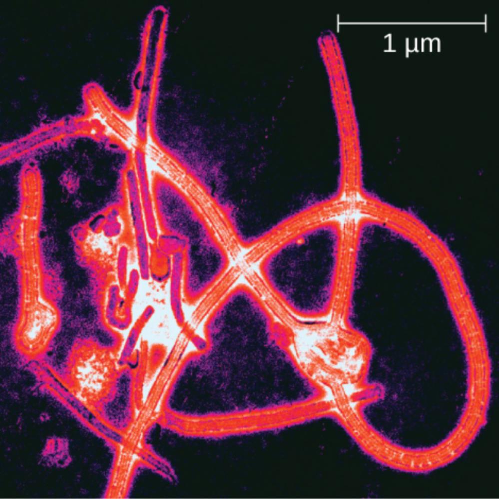
1 µm: This scale bar represents a length of one micrometer, serving as a critical reference for understanding the size of the viral particles. While the diameter of the Ebolavirus is generally uniform at approximately 80 nanometers, this scale demonstrates the variable and often extensive length of the virions, which can stretch up to 14,000 nanometers and frequently fold back on themselves.
The Unique Structure of Filoviruses
The Ebolavirus is the most notorious member of the Filoviridae family, a classification of viruses named after the Latin word “filum,” meaning thread. As seen in the electron micrograph, these viruses are not spherical like influenza or coronaviruses but instead appear as long, twisted filaments. These filaments can take on various shapes, including U-shapes, circular forms, or the classic “shepherd’s crook” appearance (a shape resembling a cane), which is a morphological hallmark of the genus. This structural plasticity is due to the flexible helical nucleocapsid contained within the viral envelope.
Morphologically, the virus is an enveloped, non-segmented, negative-sense RNA virus. The outer envelope is derived from the host cell’s membrane and is studded with viral glycoproteins (GP). These glycoproteins appear as short spikes on the surface of the filament and play a pivotal role in the infection process. They act as the key that unlocks host cells, facilitating attachment and fusion, which allows the viral genetic material to enter the cytoplasm and begin replication.
The discovery of this distinct morphology was a significant milestone in virology, separating Ebola and Marburg viruses from other hemorrhagic fever agents. The visual identification of these thread-like structures in patient samples remains a definitive method for confirming diagnosis in high-containment laboratories. Understanding the physical structure is not just academic; the surface glycoproteins are the primary targets for neutralizing antibodies and are the basis for recent vaccine developments.
Key characteristics of the Ebolavirus include:
- Viral Family: Filoviridae (characterized by filamentous, thread-like virions).
- Genome: Single-stranded, negative-sense RNA.
- Morphology: Polymorphic, often appearing as long filaments, branched shapes, or “6” and “U” shapes.
- Surface Structure: The envelope is covered in 7-10 nm long glycoprotein spikes essential for receptor binding.
Pathogenesis and Clinical Presentation of Ebola Virus Disease
Ebola Virus Disease (EVD), formerly known as Ebola hemorrhagic fever, is a rare but severe illness in humans. The pathogenesis begins when the virus enters the body through mucous membranes or breaks in the skin. The virus primarily targets immune cells, specifically macrophages and dendritic cells. By infecting these sentinels of the immune system, Ebola effectively disables the body’s initial defense mechanisms. It inhibits the production of interferon, a protein that cells release to stop viral replication, allowing the virus to spread unchecked to lymph nodes, the liver, and the spleen.
As the infection progresses, it triggers a systemic inflammatory response often referred to as a “cytokine storm.” This excessive release of inflammatory signals leads to the impairment of the vascular system. The virus damages the endothelial cells lining the blood vessels, causing them to become leaky. Simultaneously, the infection disrupts the body’s ability to clot blood effectively (coagulopathy). This combination of vascular leakage and clotting failure results in the bleeding manifestations associated with the disease, although massive external bleeding is less common than internal fluid loss and shock.
Clinically, the incubation period can range from 2 to 21 days. The onset is typically sudden, characterized by fever, fatigue, muscle pain, headache, and sore throat. This is followed by vomiting, diarrhea, rash, symptoms of impaired kidney and liver function, and in some cases, both internal and external bleeding (e.g., oozing from gums or blood in stool). The high mortality rate, which can range from 25% to 90% depending on the strain and medical care, is usually due to severe dehydration, electrolyte imbalances, and multi-organ failure caused by shock.
Transmission and Prevention
Understanding the transmission dynamics is critical for controlling outbreaks. Ebolavirus is introduced into the human population through close contact with the blood, secretions, organs, or other bodily fluids of infected animals such as fruit bats, chimpanzees, or gorillas. Once a human is infected, person-to-person transmission occurs via direct contact with the blood or body fluids of a person who is sick with or has died from Ebolavirus. It is not an airborne virus; transmission requires direct physical contact with infectious fluids or contaminated surfaces (fomites).
This mode of transmission places healthcare workers and family members of patients at the highest risk. Strict infection control protocols, including the use of personal protective equipment (PPE) and isolation of infected patients, are the cornerstones of outbreak management. Furthermore, the virus can persist in certain body fluids, such as semen and ocular fluid, for months after recovery, necessitating long-term follow-up for survivors.
Conclusion
The terrifying efficiency of the Ebolavirus is masked by its simple, thread-like appearance under the microscope. This image captures a pathogen that has challenged global health systems and spurred rapid advancements in medical countermeasures. Through a combination of supportive care, novel therapeutics, and vaccination, the medical community continues to improve survival rates, transforming a once-mysterious filovirus into a manageable, albeit still dangerous, public health threat.

