Taenia saginata, widely recognized as the beef tapeworm, is a significant zoonotic parasite that inhabits the human intestinal tract. This large ribbon-like flatworm is the causative agent of taeniasis in humans, a condition resulting from the consumption of raw or undercooked beef containing infective larval cysts. Understanding the anatomy and lifecycle of this cestode is vital for medical professionals and public health officials working to control parasitic infections and ensure food safety standards.
Overview of Taenia Saginata Morphology and Ecology
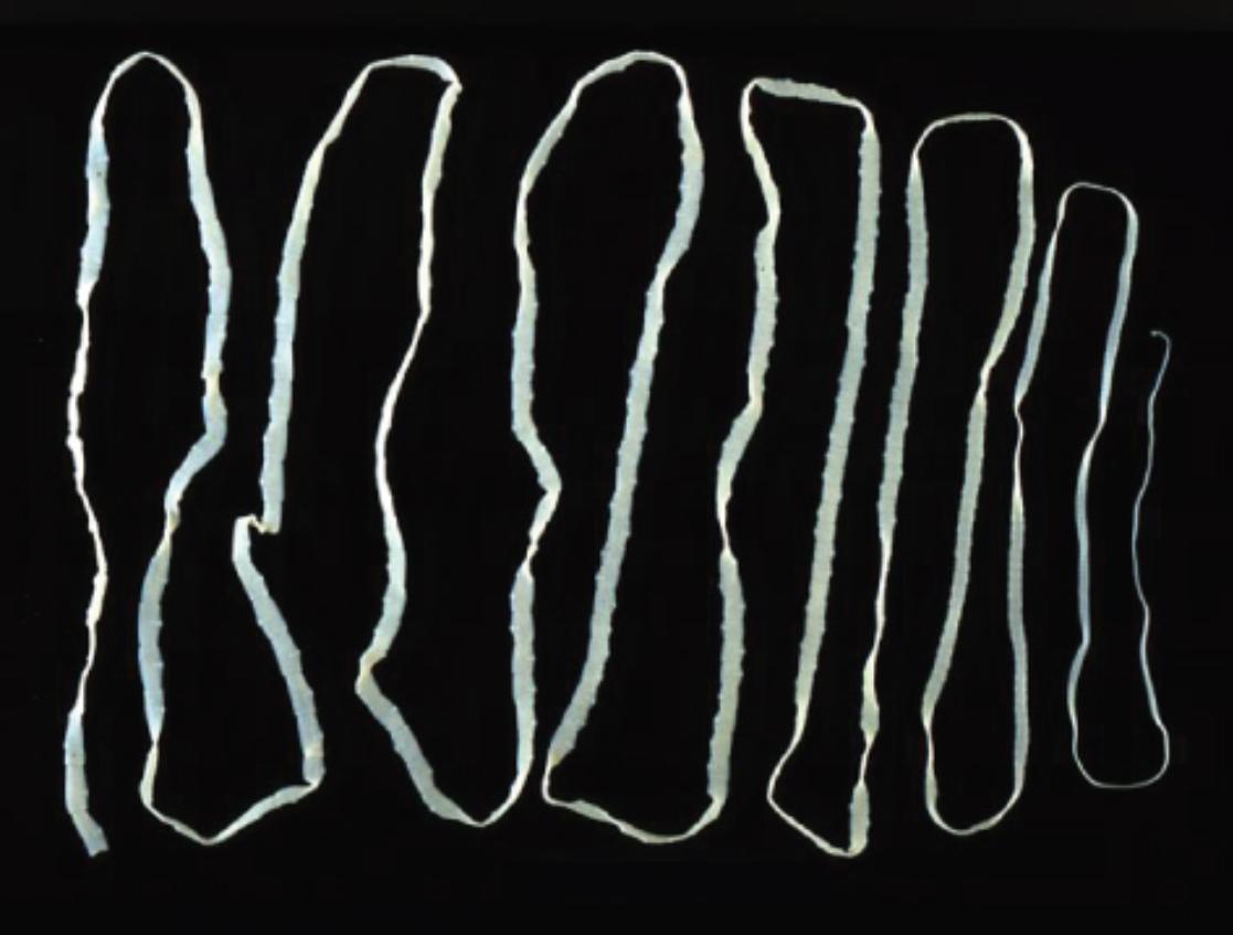
The image provided presents a striking visual of an adult Taenia saginata, a parasitic platyhelminth belonging to the class Cestoda. Unlike microscopic pathogens, the adult beef tapeworm is macroscopically immense, capable of reaching lengths between 4 to 10 meters, and in rare instances, exceeding 25 meters. The organism lacks a digestive system of its own; instead, it resides in the small intestine of the human host, absorbing predigested nutrients directly through its specialized outer skin, known as the tegument. This parasitic relationship can persist for years if left untreated, with the worm residing quietly within the gut lumen.
The lifecycle of Taenia saginata is complex and requires two distinct hosts to complete its development. Humans serve as the definitive host, harboring the sexually mature adult worm, while cattle act as the intermediate host. The infection cycle perpetuates when cattle ingest vegetation contaminated with human feces containing tapeworm eggs or gravid segments. Once inside the cow, the eggs hatch into oncospheres, migrate through the intestinal wall, and encyst in the muscle tissue, forming cysticerci. Humans become infected by eating this “measly beef” if it has not been cooked sufficiently to kill the larvae.
While the sheer size of the worm depicted in the image is alarming, the infection is often less medically severe than that of its close relative, Taenia solium (the pork tapeworm). This is primarily because T. saginata does not cause cysticercosis in humans—a condition where larvae invade human tissues such as the brain. However, the beef tapeworm remains a global health burden, particularly in regions where cattle husbandry is common and sanitation infrastructure is limited.
Key biological characteristics of Taenia saginata include:
- Scolex (Head): Possesses four powerful suckers for attachment but lacks the rostellum and hooks seen in pork tapeworms.
- Strobila: The body consists of a chain of 1,000 to 2,000 segments called proglottids.
- Reproduction: It is hermaphroditic, with each mature proglottid containing both male and female reproductive organs.
- Motility: Detached gravid proglottids are actively motile and can often force their way out of the anal sphincter.
Anatomical Structure and Physiology
The structural integrity of T. saginata is adapted perfectly for a parasitic existence. The anterior end, or scolex, anchors the worm to the mucosal wall of the jejunum. Behind the scolex lies a short neck region, which is the zone of proliferation. Here, new segments are continuously generated, pushing older segments toward the posterior end. As these segments mature, they become filled with thousands of eggs.
The most distinct feature of T. saginata, distinguishing it from other tapeworms, is the structure of its gravid proglottids. These segments are longer than they are wide and contain a highly branched uterus (typically 15 to 20 lateral branches on each side). When these segments become fully gravid, they detach from the main body (the strobila) and are passed in the stool. Uniquely, these segments are capable of independent movement and may migrate out of the anus, causing significant psychological distress and physical itching for the patient.
Clinical Presentation: Taeniasis
The infection caused by the adult worm is known as taeniasis. In many clinical cases, the infection is asymptomatic or presents with mild, non-specific gastrointestinal symptoms. However, as the worm grows and consumes nutrients intended for the host, patients may experience abdominal pain, nausea, digestive disturbances, and weight loss despite a normal appetite. The most common complaint leading to diagnosis is the observation of mobile, white segments in the feces or on undergarments.
While complications are rare, the sheer physical bulk of the worm can occasionally lead to acute issues. A large tangle of worms can cause intestinal obstruction, and individual proglottids have been known to migrate into the appendix or bile duct, causing appendicitis or cholangitis, respectively. Unlike the pork tapeworm, T. saginata poses no risk of autoinfection resulting in tissue cysts (cysticercosis) in humans; its pathogenicity is strictly limited to the intestinal lumen.
Diagnosis is confirmed through the microscopic examination of stool samples to identify eggs or by analyzing the morphology of the expelled proglottids. Treatment is generally straightforward and highly effective. Anthelmintic medications, such as Praziquantel or Niclosamide, are the drugs of choice. These medications work by either paralyzing the worm, causing it to release its grip on the intestinal wall, or by inhibiting its energy metabolism, leading to its digestion and expulsion.
Conclusion
The image of Taenia saginata serves as a potent reminder of the biological complexity of parasitic organisms and the importance of food safety. While modern medicine provides effective treatments for taeniasis, prevention remains the primary strategy. This includes adequate cooking of beef products to an internal temperature of at least 145°F (63°C) and maintaining proper sanitation to prevent the contamination of livestock grazing areas. Healthcare providers must remain vigilant in recognizing the signs of this parasitic infection to prevent long-term morbidity and interrupt the transmission cycle.

