Giardia lamblia is a microscopic protozoan parasite responsible for one of the most common waterborne diseases worldwide, known as giardiasis. This scanning electron micrograph captures the organism in its active trophozoite stage, revealing the distinct structural adaptations that allow it to colonize the human small intestine and disrupt nutrient absorption. Understanding the morphology and behavior of this pathogen is essential for healthcare providers when diagnosing gastrointestinal distress and implementing public health strategies to prevent transmission.
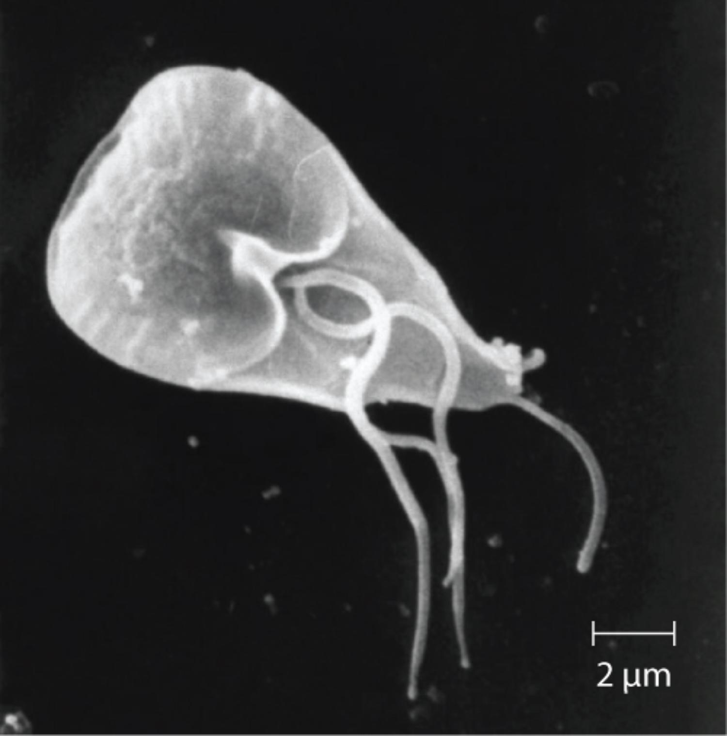
2 μm Scale Bar: This graphical indicator represents a length of two micrometers, emphasizing the microscopic nature of the organism. Since the trophozoite depicted is roughly 5 to 7 times the length of this bar, it aligns with the standard biological size of Giardia lamblia, which typically measures between 10 and 15 micrometers in length.
Understanding the Morphology of Giardia Lamblia
The image displays Giardia lamblia in its trophozoite form, the active, motile stage of its life cycle that resides within the host’s digestive tract. Structurally, the organism is characterized by a pear-shaped (pyriform) body and bilateral symmetry. A defining feature visible in high-resolution microscopy is the presence of multiple flagella—whip-like appendages that extend from the cell body. These structures are critical for motility, allowing the parasite to swim vigorously through the mucus layer of the small intestine to reach the epithelial cells where colonization occurs.
Physiologically, the ventral side of the trophozoite—the side facing the intestinal wall—features a large, specialized adhesive disc (often called a sucking disc). While not fully visible from the dorsal angle shown in some micrographs, this disc acts like a suction cup. It creates a vacuum that allows the parasite to attach firmly to the microvilli of the duodenal and jejunal epithelium. This attachment mechanism is vital for the parasite’s survival, preventing it from being swept away by peristalsis (the natural muscular contractions of the gut).
The life cycle of Giardia alternates between this fragile trophozoite stage and a robust cyst stage. While the trophozoite causes the symptoms of the disease, it cannot survive long outside the body. Conversely, the cyst form is encased in a protective shell, allowing it to withstand harsh environmental conditions, such as cold water and chlorination. Infection begins when a host ingests these dormant cysts, which then excyst in the small intestine, releasing the active trophozoites to multiply.
Key characteristics of the Giardia lamblia trophozoite include:
- Shape: Distinctive pear or teardrop shape with a rounded anterior and pointed posterior.
- Motility: Movement is facilitated by four pairs of flagella (anterior, posterior, ventral, and caudal).
- Nuclei: It possesses two nuclei, giving it a characteristic “face-like” appearance under light microscopy.
- Habitat: It primarily colonizes the duodenum and jejunum of the small intestine.
Clinical Presentation and Pathophysiology of Giardiasis
The infection caused by this parasite, Giardiasis, manifests primarily as a gastrointestinal illness. Unlike some other parasites that invade the intestinal tissue or enter the bloodstream, Giardia lamblia generally remains luminal. Its pathogenicity is largely due to the mechanical blockade it creates. By carpeting the intestinal wall with their adhesive discs, the trophozoites damage the microvilli, leading to enzyme deficiencies (such as temporary lactose intolerance) and significant malabsorption of fats and carbohydrates.
Clinically, the hallmark of giardiasis is diarrhea, which can be acute or chronic. The stool is often described as “steatorrhea,” meaning it is greasy, foul-smelling, and floats, indicating the body’s inability to absorb fats properly. Other common symptoms include abdominal cramping, bloating, excessive gas (flatulence), nausea, and weight loss. In children, chronic infection is particularly concerning as severe malabsorption can lead to failure to thrive and delayed physical development.
Diagnosis is typically confirmed through stool antigen tests, which are more sensitive than traditional microscopic examination of ova and parasites. Once diagnosed, the infection is generally treatable with antimicrobial agents such as Metronidazole, Tinidazole, or Nitazoxanide. Prevention focuses on breaking the transmission cycle, which is primarily fecal-oral. This involves rigorous hand hygiene, filtering or boiling water from natural sources like lakes or streams, and proper sanitation practices in communal settings like daycare centers.
Conclusion
The electron micrograph of Giardia lamblia offers a detailed view of a pathogen that has evolved highly effective mechanisms for parasitic success. By utilizing its flagella for navigation and its adhesive disc for attachment, this trophozoite can significantly disrupt the digestive processes of its host. Recognizing the specific morphology of Giardia not only aids in clinical identification but also highlights the complex biological interactions between human hosts and the microscopic world of intestinal parasites.

