Mechanical heart valves are durable prosthetic devices designed to replicate the critical function of native heart valves in patients suffering from severe valvular disease or dysfunction. Unlike biological valves derived from animal tissue, these mechanical counterparts are engineered from robust materials like titanium and pyrolytic carbon, offering longevity that can last a patient’s lifetime. The image above displays a historical collection of these devices, illustrating the technological progression from early ball-and-cage models to modern bileaflet designs, each created to optimize blood flow and reduce complications.
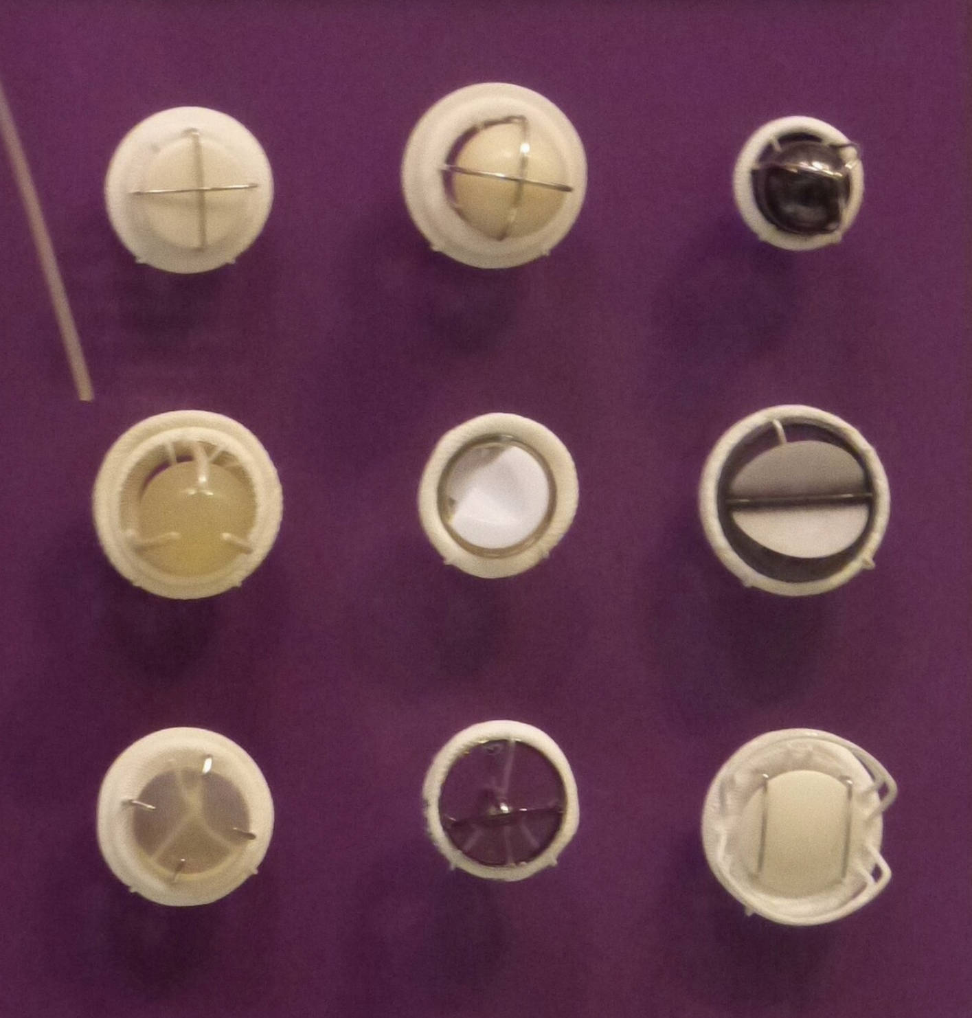
Ball-and-Cage Valve: This early design, visible in the top row, consists of a silicone ball housed within a metal cage. When blood pressure builds, the ball moves forward to allow flow, and when pressure drops, it falls back into the ring to seal the valve, though its bulky profile often altered blood flow dynamics.
Tilting Disc Valve: Seen in the middle variations, this design utilizes a single circular disc held in place by metal struts. The disc tilts open to an angle to allow blood to pass and tilts back to close the orifice, offering better hemodynamics and a lower profile than the ball-and-cage models.
Bileaflet Valve: Visible in the collection, this represents the modern standard for mechanical heart valves, featuring two semicircular leaflets that pivot on hinges. This design creates a central flow opening that closely mimics natural physiology, significantly reducing turbulence and the work required by the heart to pump blood.
Engineering the Human Heartbeat
The image provided showcases a variety of mechanical heart valves housed in the National Museum of Health and Medicine, representing decades of cardiovascular innovation. The human heart contains four valves—the tricuspid, pulmonary, mitral, and aortic—that act as one-way gates to ensure blood flows in the correct direction. When these valves become diseased, they can either become too narrow (stenosis) or fail to close properly (regurgitation), placing immense strain on the heart muscle.
Surgical replacement becomes necessary when valve repair is not feasible. Mechanical valves are often preferred for younger patients because of their exceptional durability. While biological valves (made from pig or cow tissue) eventually wear out and require replacement after 10 to 15 years, a mechanical valve is structurally designed to last indefinitely. However, the engineering challenge has always been to create a device that minimizes damage to blood cells and prevents the formation of blood clots.
The evolution from the early ball-and-cage models to the sophisticated bileaflet designs shown in the image highlights the medical community’s pursuit of the perfect hemodynamic profile. Early valves were effective but bulky, often causing turbulence in the blood flow. Newer designs have focused on maximizing the effective orifice area—the actual space available for blood to flow through—thereby reducing the pressure gradient across the valve.
Key considerations for mechanical heart valve therapy include:
- Durability: They are built to withstand the millions of beats a heart performs annually without structural failure.
- Auditory Feedback: Patients often report hearing a soft “clicking” sound as the mechanical components close, which is a normal characteristic of the device.
- Thrombogenicity: Because the materials are artificial, blood is more prone to clotting on the surface, necessitating lifelong medical management.
Valvular Heart Disease and Surgical Intervention
The primary medical indication for implanting the devices shown in the image is valvular heart disease. This condition can be congenital, meaning a person is born with a malformed valve, or acquired through infections like rheumatic fever, age-related calcification, or endocarditis. In aortic stenosis, for example, the aortic valve leaflets become stiff and calcified, preventing the valve from opening fully. This forces the left ventricle to pump harder to eject blood into the aorta, leading to hypertrophy (thickening) of the heart muscle and eventual heart failure if left untreated.
Conversely, in valvular regurgitation, the valve leaflets fail to close completely. This causes blood to leak backward into the heart chamber from which it was just pumped. In severe mitral regurgitation, blood flows back into the left atrium during contraction, causing fluid backup in the lungs (pulmonary edema) and shortness of breath. Mechanical valves restore normal hemodynamics by providing a competent seal and a wide opening for blood flow, effectively relieving the strain on the heart.
Life with a Mechanical Valve: Anticoagulation
While the structural integrity of mechanical valves is superior to biological options, they present a unique physiological challenge: the risk of thrombosis (clot formation). The artificial surfaces of the valve (carbon, metal, and polyester sewing rings) can trigger the body’s clotting cascade. If a clot forms on the valve, it can obstruct the mechanism or break loose and travel to the brain, causing a stroke.
To prevent this, patients with mechanical heart valves must undergo lifelong anticoagulation therapy, typically with a medication called warfarin (Coumadin). This requires regular blood monitoring to ensure the International Normalized Ratio (INR) remains within a specific therapeutic range—thin enough to prevent clots but thick enough to prevent excessive bleeding. Despite this requirement, the trade-off is often acceptable for patients who wish to avoid the risks associated with re-operation that come with tissue valves.
Conclusion
The array of mechanical heart valves featured in the image stands as a testament to the ingenuity of biomedical engineering. From the robust simplicity of the ball-and-cage to the streamlined efficiency of the bileaflet valve, these devices have extended and improved the lives of countless individuals with heart disease. As technology continues to advance, future iterations aim to reduce the need for aggressive blood-thinning medication while maintaining the legendary durability that characterizes these life-saving prosthetics.

