Introduction to Pediatric Thoracic Imaging
Pediatric thoracic X-rays are crucial diagnostic tools in assessing respiratory conditions in children. Interpreting these images requires a comprehensive understanding of normal pediatric anatomy and common pathologies. This article will delve into a case study, focusing on the interpretation of a chest X-ray and the associated clinical presentation and management.
Clinical Presentation
The patient is a male infant with a stable general condition and vital signs.
Physical Examination Findings:
- Skin: Natural, no rashes observed.
- Head and Neck: No cervical lymphadenopathy.
- Oropharynx: Normal.
- Respiratory System: The patient exhibits inspiratory and expiratory rhonchi and crackles, described as minimal rales. Eupneic, being monitored with a reservoir mask, with oxygen saturation (SpO2) at 98%.
- Cardiovascular System: Normal heart sounds (S1, S2) with no additional sounds or murmurs. Peripheral pulses are palpable bilaterally.
- Gastrointestinal System: Abdomen is soft, non-tender, with no defense or rebound tenderness. Bowel sounds present, with normal gas and stool passage.
- Genitourinary System: Normal male genitalia, with adequate urine output.
- Neuromuscular System: Glasgow Coma Scale (GCS) of 15. Pupils are isochoric and reactive to light. Neuromotor development is appropriate for age. No signs of meningeal irritation.
- Extremities: No deformities noted.
Radiological Findings: Chest X-ray
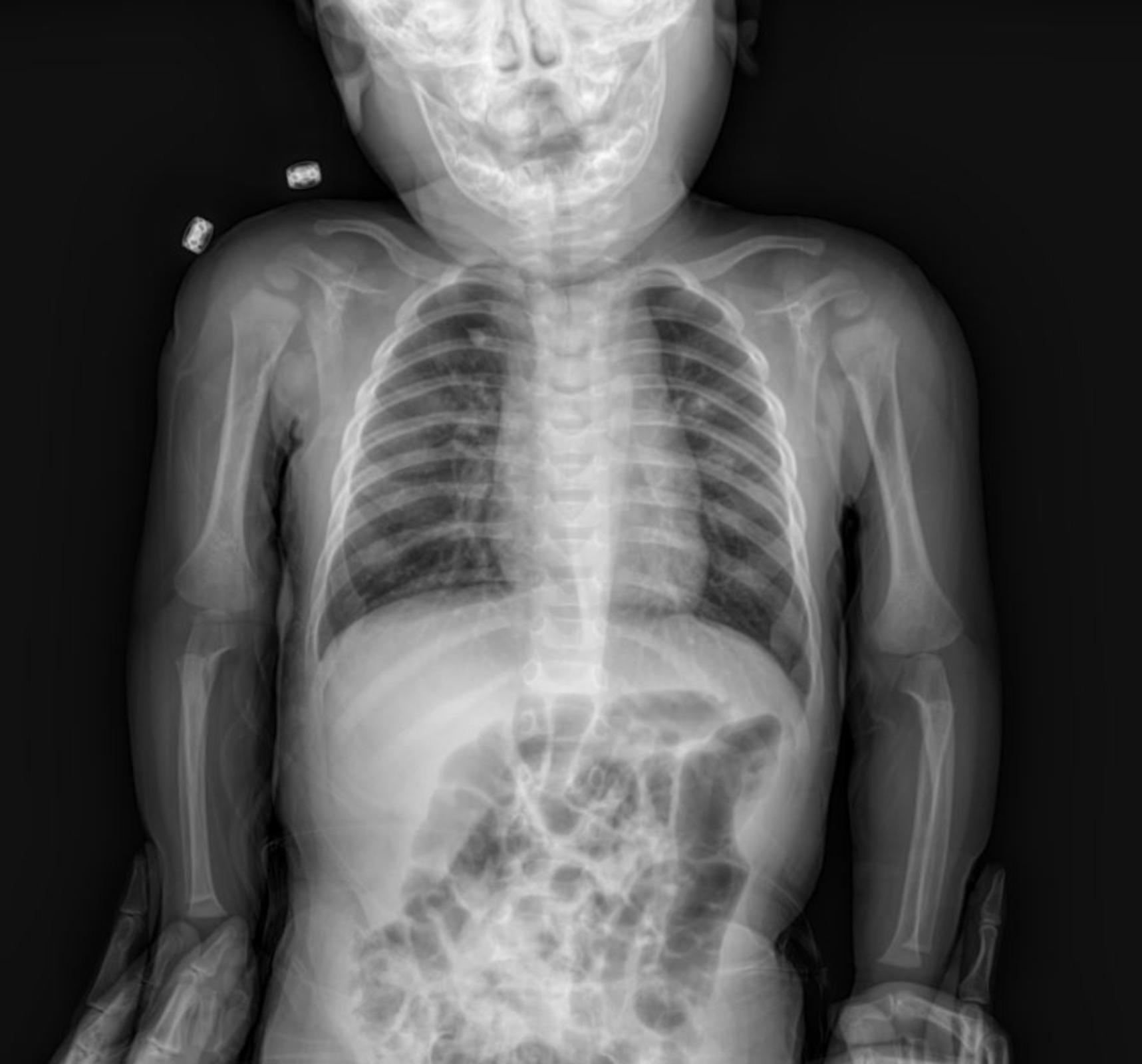
The provided anterior-posterior (AP) chest X-ray of the infant reveals several key features:
- Lungs: The lung fields appear adequately inflated. There is evidence of increased bronchovascular markings, particularly in the perihilar regions, which can suggest bronchial wall thickening or fluid accumulation. Additionally, subtle paracardial infiltration is noted, indicating a mild inflammatory or infectious process adjacent to the heart borders.
- Heart: The cardiac silhouette appears within normal limits for size and contour for a pediatric patient, without overt cardiomegaly. The paracardial infiltration, while present, does not appear to significantly distort the cardiac borders.
- Diaphragm: The diaphragmatic contours are clear and sharp, with no evidence of pleural effusions.
- Bony Thorax: The ribs and clavicles appear intact with no obvious fractures or deformities. The skeletal structures are appropriate for the child’s age.
- Trachea: The trachea appears patent and centrally located.
- Foreign Bodies: Two metallic densities are noted projected over the left upper chest/shoulder region. These would need clinical correlation to determine their nature (e.g., external artifacts, monitoring leads).
Given the clinical presentation of rhonchi and rales, combined with the X-ray findings of increased bronchovascular markings and mild paracardial infiltration, the overall picture is strongly suggestive of a lower respiratory tract infection, such as bronchiolitis or pneumonia, common in infants. The paracardial infiltration further supports an inflammatory process within the lung parenchyma in this region.
Management Plan
The patient is currently receiving the following treatments:
- Bronchodilators:
- Salbutamol nebulization with proper doses.
- Ipratropium bromide nebulization with proper doses.
- Corticosteroids:
- Budesonide nebulization with proper doses.
- Prednisolone intravenous administration with proper doses.
- Other Medications:
- Magnesium sulfate intravenous administration with proper doses (diluted in 30 mL normal saline).
- Macrolides oral administration with proper doses.
- Ampicillin intravenous administration with proper doses.
Disclaimer: Drug types and dosages should always be confirmed with the relevant dosage guidelines in your country or by consulting a licensed physician in the department. This information is for educational purposes only.
Conclusion
This case highlights the integration of clinical findings with radiological interpretation in the diagnosis and management of pediatric respiratory conditions. The chest X-ray provided valuable insights into the status of the lungs and surrounding structures, guiding the therapeutic approach.
Please note: This content is intended for educational purposes and reference only. It should not be used as a substitute for professional medical advice, diagnosis, or treatment. Always seek the advice of a qualified healthcare provider for any medical concerns.

