This chest X-ray (CXR) diagram illustrates dilated cardiomyopathy, a serious condition affecting the heart’s ability to pump blood effectively. The image provides a clear visual representation of cardiac enlargement, which is a hallmark feature of this disease. By examining such radiological findings, medical professionals can gain crucial insights into the extent of heart remodeling and guide appropriate diagnostic and therapeutic strategies for patients suffering from this condition.
Introduction to Dilated Cardiomyopathy on Chest X-Ray
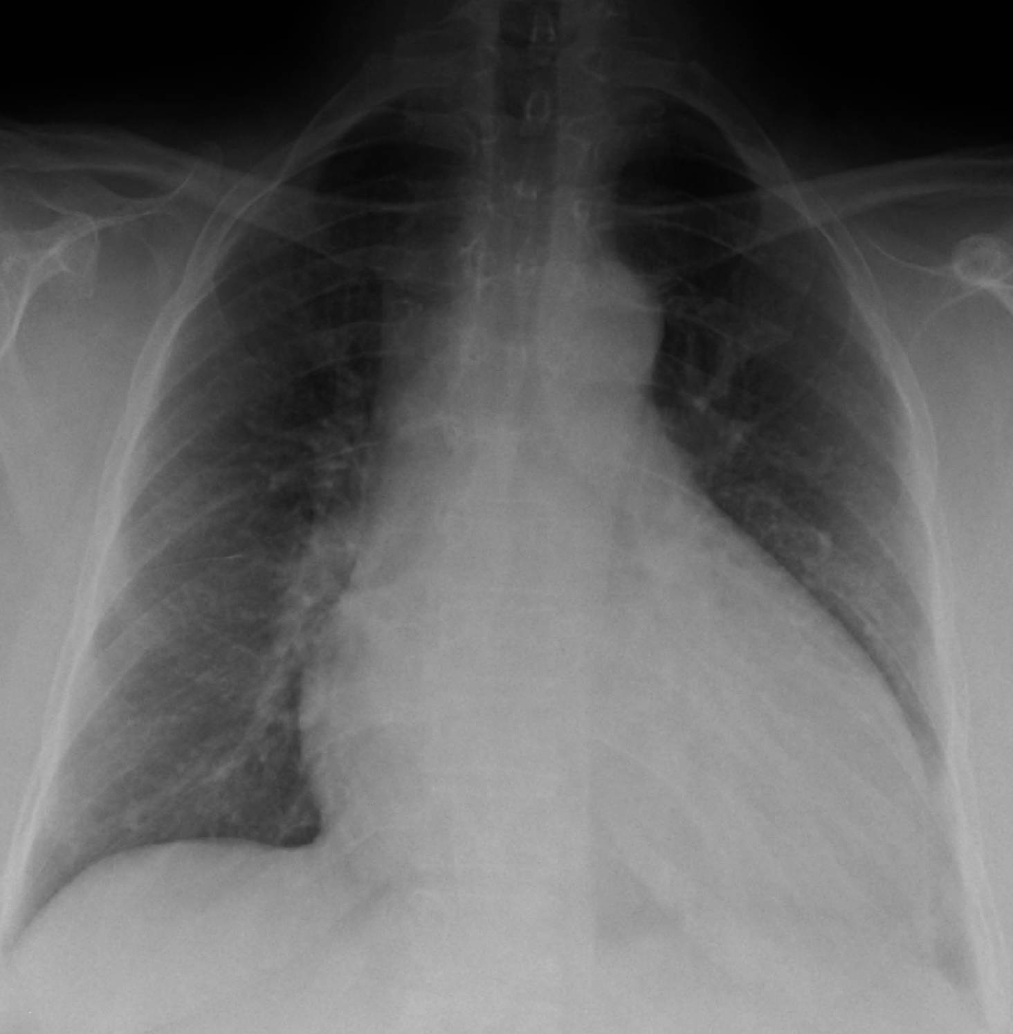
Dilated cardiomyopathy (DCM) is a type of heart muscle disease characterized by the enlargement and weakening of the heart’s main pumping chamber, the left ventricle. This progressive condition impairs the heart’s ability to pump blood efficiently to the rest of the body, leading to symptoms of heart failure. A chest X-ray, as depicted here, is often one of the initial diagnostic tools used to identify significant cardiac enlargement, a key indicator of DCM. While not definitive on its own, a CXR provides valuable clues that prompt further, more specific investigations.
The image vividly demonstrates the hallmark features of dilated cardiomyopathy: an enlarged cardiac silhouette. This enlargement is a direct consequence of the ventricular dilation and thinning of the heart muscle walls that occur in DCM. Understanding these radiological signs is crucial for early detection and for guiding subsequent diagnostic steps, which typically include echocardiography for functional assessment and other advanced imaging or genetic tests to determine the underlying cause.
DCM can arise from a multitude of factors, ranging from genetic predispositions to environmental insults. Its impact extends beyond the heart, potentially affecting other organ systems due to reduced cardiac output. Early identification of the characteristic cardiac changes on a chest X-ray is therefore essential for prompt management and to improve patient outcomes.
Several factors can contribute to the development of dilated cardiomyopathy:
- Genetic Factors: A significant percentage of DCM cases are hereditary, with mutations in genes encoding for sarcomeric proteins or other structural components of the heart.
- Infections: Viral infections, particularly myocarditis (inflammation of the heart muscle), can sometimes lead to DCM.
- Toxins: Chronic alcohol abuse and certain chemotherapy drugs (e.g., anthracyclines) are known to be cardiotoxic and can induce DCM.
- Ischemic Heart Disease: While DCM is distinct from ischemic heart disease, prolonged untreated ischemia can lead to ischemic cardiomyopathy, which shares features with DCM.
- Autoimmune Diseases: Conditions like lupus or rheumatoid arthritis can sometimes contribute to myocardial damage.
- Metabolic Disorders: Uncontrolled diabetes or thyroid dysfunction can also play a role in the development of DCM.
- Peripartum Cardiomyopathy: A rare form of DCM that develops during late pregnancy or in the months following childbirth.
Identifying the underlying cause is paramount for targeted treatment and management of the disease.
The Pathophysiology of Dilated Cardiomyopathy
Dilated cardiomyopathy primarily affects the heart’s ventricles, particularly the left ventricle, causing them to stretch and thin. This dilation leads to an increase in the chamber volume but a decrease in the heart’s ability to contract forcefully, a condition known as systolic dysfunction. As the heart attempts to compensate for this reduced pumping efficiency, it undergoes a process called remodeling, where the muscle walls stretch further, and the chambers enlarge even more. This compensatory mechanism is ultimately detrimental, as it increases the wall stress and oxygen demand of the already weakened heart.
The ineffective pumping of blood results in reduced cardiac output, meaning less oxygenated blood reaches the body’s tissues and organs. Simultaneously, blood can back up in the pulmonary veins and lungs, leading to pulmonary congestion and symptoms like shortness of breath. Over time, the enlargement of the heart chambers can also stretch the mitral and tricuspid valves, leading to regurgitation (leaky valves), which further exacerbates the heart’s inefficiency. The chronic strain on the heart muscle can also predispose patients to arrhythmias, including potentially life-threatening ventricular tachycardias or fibrillation.
Radiological Features of Dilated Cardiomyopathy on CXR
A chest X-ray in a patient with dilated cardiomyopathy typically reveals several characteristic findings. The most prominent feature, as illustrated in the diagram, is cardiomegaly, which refers to an enlarged heart silhouette. The cardiothoracic ratio (the ratio of the maximum horizontal cardiac diameter to the maximum horizontal thoracic diameter) is often increased, usually exceeding 0.5. The heart appears globular or “pear-shaped” due to the significant dilation of all four cardiac chambers, though the left ventricle is typically the most affected and contributes most to the enlargement seen on a PA (posteroanterior) view.
In addition to cardiomegaly, signs of pulmonary venous hypertension and pulmonary edema may also be evident, reflecting the backward flow of blood due to the heart’s impaired pumping ability. These signs can include:
- Cephalization of pulmonary vessels: The upper lobe pulmonary veins appear engorged as blood is shunted upwards due to increased pressure.
- Kerley B lines: Small, horizontal lines seen at the lung periphery, indicating interstitial edema.
- Pleural effusions: Fluid accumulation in the space between the lungs and the chest wall, often seen as blunting of the costophrenic angles.
While a CXR can provide compelling evidence for DCM, it cannot definitively diagnose the condition or precisely quantify cardiac function. It serves as an excellent screening tool, prompting further diagnostic tests such as echocardiography, which provides detailed images of heart structure and function, including ejection fraction, a critical measure of pumping efficiency.
Management and Prognosis of Dilated Cardiomyopathy
The management of dilated cardiomyopathy is multifaceted and aims to improve cardiac function, alleviate symptoms, prevent complications, and enhance quality of life. Treatment strategies often involve a combination of medications, lifestyle modifications, and in some cases, advanced therapies. Medications commonly prescribed include ACE inhibitors or ARBs, beta-blockers, and mineralocorticoid receptor antagonists, all of which help to reduce the workload on the heart, improve its pumping ability, and prevent further remodeling. Diuretics are often used to manage fluid retention and relieve symptoms of congestion.
Lifestyle modifications, such as adhering to a low-sodium diet, limiting fluid intake, avoiding alcohol, and engaging in appropriate physical activity, are crucial supportive measures. For patients with severe DCM and persistent symptoms, advanced therapies may be considered. These include implantable cardioverter-defibrillators (ICDs) to prevent sudden cardiac death from arrhythmias, cardiac resynchronization therapy (CRT) for specific conduction abnormalities, and in end-stage cases, left ventricular assist devices (LVADs) or heart transplantation. Regular monitoring and follow-up are essential to adjust treatment as the disease progresses and to manage potential complications, ultimately striving to optimize the patient’s long-term outlook.

