The alimentary canal, the muscular tube extending from the esophagus to the anus, is a marvel of biological engineering. Understanding its intricate layers is fundamental to grasping how digestion, absorption, and protection occur within the human body. This article delves into the four primary tissue layers—mucosa, submucosa, muscularis, and serosa—and their specialized components, offering a detailed look at the structural foundation of the digestive system.
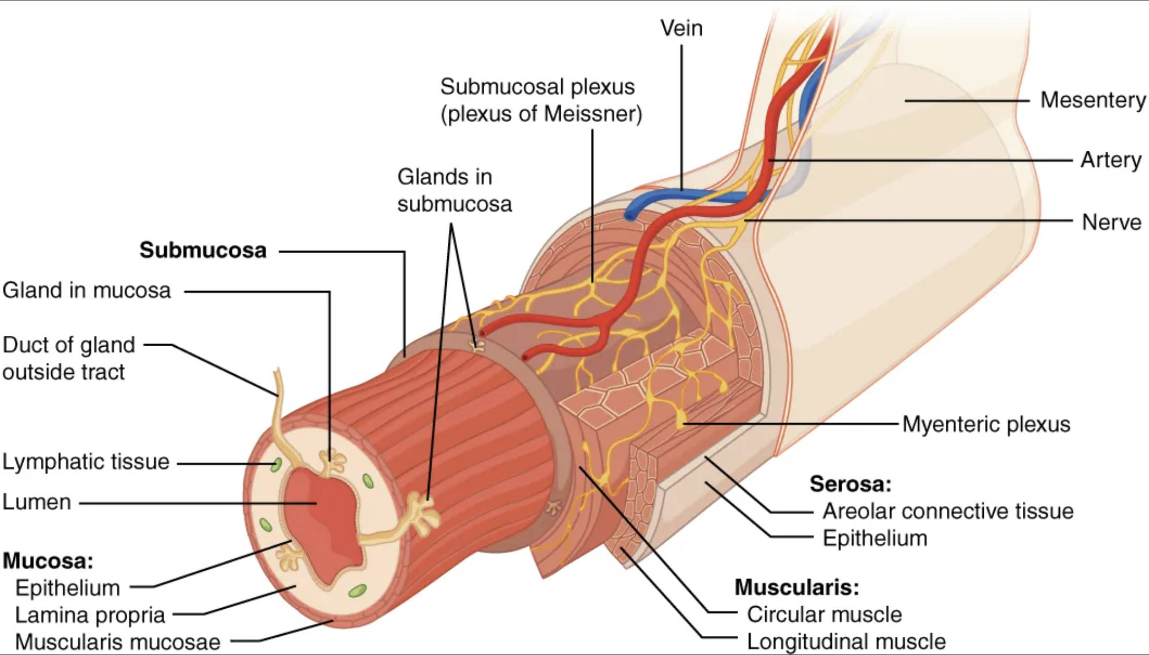
Mesentery: This double layer of peritoneum anchors the alimentary canal to the posterior abdominal wall. It provides a pathway for blood vessels, nerves, and lymphatic vessels to reach the digestive organs, supporting their metabolic and regulatory needs.
Artery: These blood vessels supply oxygenated blood and nutrients to the various layers of the alimentary canal. They are crucial for the high metabolic demands of the digestive tissues, facilitating processes like secretion, absorption, and muscle contraction.
Vein: These blood vessels carry deoxygenated blood and absorbed nutrients away from the alimentary canal. They are vital for transporting digested food components from the small intestine to the liver and then to the rest of the body.
Nerve: These neural pathways, part of the enteric nervous system and extrinsic innervation, regulate the functions of the alimentary canal. They control muscle contractions (peristalsis), glandular secretions, and local blood flow within the digestive tract.
Submucosal plexus (plexus of Meissner): Located within the submucosa, this network of nerves is part of the enteric nervous system. It primarily regulates glandular secretions in the mucosa and controls local blood flow, influencing the chemical aspects of digestion.
Glands in submucosa: These glands are embedded within the submucosal layer and produce various secretions, such as mucus and enzymes. Their ducts typically extend into the lumen, contributing to lubrication and chemical digestion.
Myenteric plexus: Situated between the circular and longitudinal muscle layers of the muscularis, this nerve plexus is crucial for controlling gastrointestinal motility. It primarily regulates the strength and frequency of muscle contractions, driving the movement of food through the canal.
Serosa: This is the outermost layer of the alimentary canal in most regions, consisting of areolar connective tissue and epithelium. It provides a protective covering and secretes serous fluid, which lubricates the outer surface of the organ, allowing it to move freely within the abdominal cavity.
Areolar connective tissue (within Serosa): This loose connective tissue forms the bulk of the serosa, providing flexibility and support. It contains blood vessels, nerves, and lymphatic vessels, contributing to the overall health and function of the digestive tract.
Epithelium (within Serosa): This simple squamous epithelium, also known as the visceral peritoneum, forms the smooth outer surface of the alimentary canal. It reduces friction as the digestive organs move against each other and the abdominal wall.
Muscularis: This layer is primarily responsible for the movements of the alimentary canal, particularly peristalsis and segmentation. It typically consists of two sub-layers of smooth muscle, which contract rhythmically to propel and mix food.
Circular muscle: This inner layer of smooth muscle within the muscularis is arranged circumferentially around the lumen. Its contractions narrow the lumen, aiding in segmentation and the forward propulsion of the food bolus.
Longitudinal muscle: This outer layer of smooth muscle within the muscularis is arranged along the length of the alimentary canal. Its contractions shorten the tube, contributing to both mixing and propulsive movements.
Submucosa: This layer lies beneath the mucosa and consists of dense irregular connective tissue. It contains blood vessels, lymphatic vessels, nerves (including the submucosal plexus), and sometimes glands, providing support and facilitating nutrient transport.
Gland in mucosa: These glands are located within the mucosal layer and are responsible for secreting mucus, digestive enzymes, and hormones directly into the lumen. They play a direct role in chemical digestion and protection of the epithelial lining.
Duct of gland outside tract: This refers to the excretory duct of a gland situated outside the wall of the alimentary canal, such as the pancreas or salivary glands. These ducts transport digestive juices into the lumen to aid in digestion.
Lymphatic tissue: This tissue, often found in the mucosa and submucosa, is a crucial component of the immune system. It protects the digestive tract from pathogens that may enter with food, forming a first line of defense.
Lumen: This is the hollow, central space within the alimentary canal through which food passes. It is where digestion and absorption primarily occur, and its size can vary depending on the organ and the presence of food.
Mucosa: This is the innermost layer of the alimentary canal, directly lining the lumen. It is specialized for secretion of mucus and digestive enzymes, and for the absorption of digested nutrients into the bloodstream.
Epithelium (within Mucosa): This innermost layer of the mucosa is in direct contact with the food. It is typically a simple columnar epithelium in most of the digestive tract, specialized for absorption and secretion, and often contains goblet cells that produce mucus.
Lamina propria: This layer of loose areolar connective tissue supports the epithelium of the mucosa. It contains capillaries for nutrient absorption, lymphatic vessels, and scattered lymphoid tissue for immune surveillance.
Muscularis mucosae: This thin layer of smooth muscle is part of the mucosa, situated between the lamina propria and the submucosa. Its contractions cause local movements of the mucosa, forming folds that increase surface area for absorption and secretion.
The alimentary canal, also known as the gastrointestinal (GI) tract, is a continuous muscular tube that extends from the mouth to the anus. Its primary function is to digest food, absorb nutrients, and eliminate waste products. This complex organ system is meticulously structured with four distinct tissue layers, each contributing uniquely to its overall function. Understanding these layers is paramount for comprehending digestive physiology and pathology.
The four fundamental layers of the alimentary canal are:
- Mucosa: The innermost layer, specialized for secretion and absorption.
- Submucosa: Contains blood vessels, nerves, and lymphatic tissue.
- Muscularis: Responsible for motility through its smooth muscle layers.
- Serosa: The outermost protective layer.
These layers work in concert to facilitate the intricate processes of digestion. From the mechanical breakdown of food to the chemical digestion by enzymes and the subsequent absorption of nutrients, each layer plays a vital role. The structural integrity and coordinated function of these layers are essential for maintaining digestive health.
The Mucosa: Interface of Digestion and Absorption
The mucosa is the innermost lining of the alimentary canal and is arguably the most dynamic layer. It is directly exposed to ingested food and plays a pivotal role in both secretion and absorption. Comprising three sub-layers—the epithelium, lamina propria, and muscularis mucosae—it is designed for maximum efficiency. The epithelial layer, typically a simple columnar epithelium in most of the GI tract, is responsible for secreting digestive enzymes and mucus, which lubricates the passage of food and protects the lining from acidic environments and abrasive food particles. The lamina propria, a connective tissue layer, supports the epithelium and contains capillaries for nutrient absorption, as well as lymphoid tissue for immune defense. Beneath these, the muscularis mucosae is a thin layer of smooth muscle that causes local movements of the mucosa, creating folds that increase surface area and enhance contact with digested food.
Submucosa and Muscularis: Support, Control, and Movement
Beneath the mucosa lies the submucosa, a layer of dense irregular connective tissue rich in blood vessels, lymphatic vessels, and nerves. This layer is crucial for nourishing the surrounding tissues and transporting absorbed nutrients. It also houses the submucosal plexus (Meissner’s plexus), a part of the enteric nervous system that regulates glandular secretions and local blood flow. The muscularis, typically composed of two layers of smooth muscle—an inner circular layer and an outer longitudinal layer—is primarily responsible for the motility of the alimentary canal. The coordinated contractions of these muscle layers produce peristalsis, wave-like contractions that propel food through the tract, and segmentation, local contractions that mix food with digestive juices. The myenteric plexus (Auerbach’s plexus), located between these muscle layers, controls these powerful contractions, ensuring efficient food movement and mixing.
The Serosa: Outer Protection and Connection
The outermost layer of the alimentary canal in the abdominal cavity is the serosa, also known as the visceral peritoneum. This thin, protective layer is composed of areolar connective tissue covered by a simple squamous epithelium. The serosa secretes a slippery serous fluid that lubricates the outer surfaces of the digestive organs, allowing them to glide smoothly against each other and against the abdominal wall during movements like peristalsis. This lubrication prevents friction and irritation, maintaining the integrity of the organs. Furthermore, the serosa is continuous with the mesentery, a double-layered peritoneal fold that anchors the digestive organs to the posterior abdominal wall and provides a pathway for blood vessels, nerves, and lymphatic vessels to reach these organs, ensuring their continued function and support.
In summary, the alimentary canal is a testament to the sophisticated organization of the human body. Each of its four layers—mucosa, submucosa, muscularis, and serosa—along with their specialized components, performs distinct yet interconnected roles that are vital for the digestion, absorption, and elimination of food. From the intricate neural networks that control movement and secretion to the protective outer coverings, every element contributes to the remarkable efficiency of the digestive system. A thorough understanding of these anatomical layers is foundational to appreciating the complex physiological processes that sustain life.

