This article presents a clinical case of a 14-year, 9-month-old female patient experiencing recurrent respiratory distress, highlighting the diagnostic approach, radiological findings, and initial management. This case provides a valuable learning opportunity for medical students and practitioners to understand the complexities of chronic respiratory issues in adolescents.
Clinical Presentation
A 14-year, 9-month-old female patient, with no known chronic illness or regular medication, presented to the emergency department on November 9, 2025, with a three-day history of cough, shortness of breath, inability to breathe particularly when lying down, and fever.
- Initial Examination Findings:
- Tachypnea
- Rales (+)
- Rhonchi (+)
- Wheezing (+)
- Retractions (+)
- Based on these findings, the patient was admitted for observation.
Radiological Findings
Given the patient’s symptoms and initial physical findings, a chest X-ray and subsequent CT scan were performed.
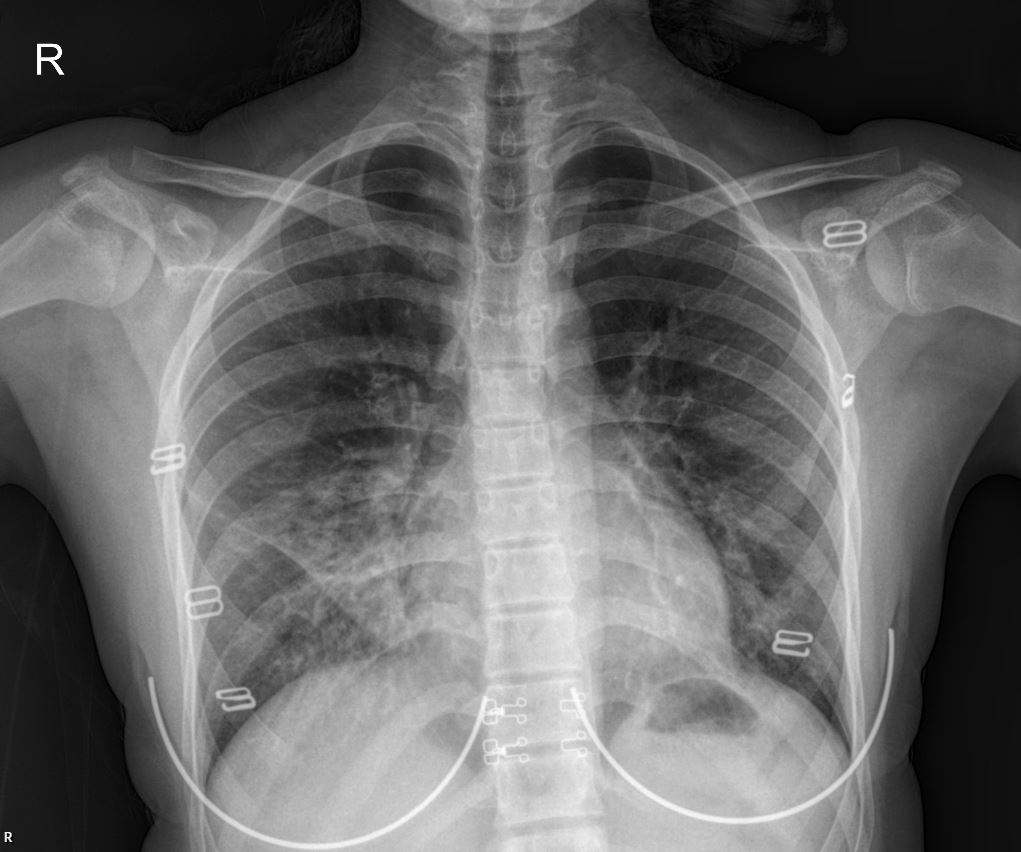
- PA Chest X-ray Interpretation: The initial PA chest X-ray (as shown above) revealed lobar involvement, indicating significant parenchymal changes consistent with an acute inflammatory process. The infiltrates are diffuse, particularly noticeable in the bilateral lower and mid lung fields, suggesting widespread involvement.
- Thoracic CT Interpretation: The thoracic CT scan further elaborated on these findings, showing “peribronchial infiltration and mild ground-glass opacities in areas of both lung parenchyma.” This suggests an inflammatory or infectious process affecting the small airways and alveoli.
Past Medical History
The patient’s history is significant for recurrent respiratory issues:
- Frequent nebulizer use over the past 5 years.
- Multiple hospital admissions due to respiratory distress.
- No prior allergy follow-up.
- The underlying cause of her recurrent respiratory distress had not been previously investigated.
- No family history of tuberculosis.
Due to her general decline and the persistent nature of her respiratory problems, the patient was admitted to the pediatrics ward for further investigation into the cause of her recurrent respiratory distress.
Current Physical Examination (On Ward)
- General condition: Moderate.
- Vitals: Stable.
- Skin: Normal, no rash.
- Head-Neck: Normal.
- Oropharynx: Normal.
- Respiratory System: Harsh inspiratory and expiratory breath sounds, coarse, crepitant rales (+), rhonchi (+). Monitored on room air, but tachypneic.
- Cardiovascular System: S1+ S2+, no extra sounds, no murmurs heard, peripheral pulses palpable bilaterally.
- Gastrointestinal System: Abdomen soft, no defense, no rebound tenderness, gas and stool passage present.
- Genitourinary System: Externally female, no major urogenital anomalies, urine output present.
- Neuromuscular System: GCS: 15, conscious, pupils isocoric, light reflex +/+, neuromotor development appropriate for age, no meningeal irritation signs.
- Extremities: Normal, no deformities.
Hospital Course (Day 1)
The patient was admitted with a preliminary diagnosis of bronchopneumonia.
Treatment Plan
- Ceftriaxone: With proper doses intravenously (Day 1).
- Ipratropium bromide: With proper doses via inhalation.
- Budesonide: With proper doses via inhalation.
- Acetylcysteine: With proper doses via nebulization.
- Methylprednisolone: With proper doses intravenously (Day 2).
Please note: Drug types and dosages should be verified according to your country’s relevant pharmaceutical dosage guidelines, or by consulting a licensed physician in the relevant department.
Future Plans
- Tuberculosis investigations are planned.
- Immunoglobulin levels will be assessed.
- Results will be closely monitored.
Conclusion
This case underscores the critical importance of investigating the underlying causes of recurrent respiratory symptoms in pediatric patients, especially when initial treatments provide only temporary relief. The combination of clinical presentation, chest X-ray findings showing lobar involvement, and CT scan results indicating peribronchial infiltration and ground-glass opacities points towards a complex respiratory pathology, potentially infectious or inflammatory. The history of frequent nebulizer use and hospital admissions highlights the need for a comprehensive diagnostic workup to identify any contributing factors, such as allergies, asthma, or immune deficiencies.
This content is for educational and reference purposes only and should not be used as a basis for diagnosis or treatment. Always consult with a qualified healthcare professional for any medical concerns.

