Witness the immersive learning experience of medical students at Monterrey Tech as they meticulously perform a heart dissection, offering a tangible insight into cardiac anatomy. This close-up view reveals the intricate internal structures, particularly the atria, providing an invaluable educational opportunity. Hands-on dissection is a cornerstone of medical education, fostering a deep understanding of human physiology and pathology.
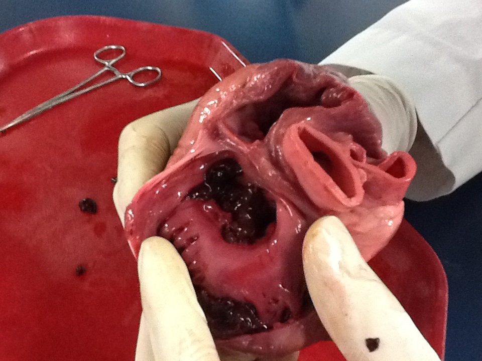
While the image does not contain explicit labels, the context provided (“entrance of the big atria can be observed”) and the visual evidence allow for an explanation of the visible anatomical features within this dissected heart specimen.
Atria (Entrance): The large, relatively thin-walled chambers at the top of the heart, visible as wide openings in the dissected specimen, are the atria. These chambers serve as receiving areas for blood returning to the heart from either the body (right atrium) or the lungs (left atrium).
Ventricular Chambers: Beneath the atria, the thicker, more muscular walls of the ventricles are visible, particularly on the left side where the wall appears substantially robust. The ventricles are the primary pumping chambers, responsible for ejecting blood out of the heart.
Cardiac Muscle (Myocardium): The predominant tissue seen throughout the dissection is the cardiac muscle, or myocardium, which appears reddish and textured. This specialized muscle tissue is responsible for the heart’s powerful and rhythmic contractions.
Heart Valves (Implied): While not perfectly in focus, the structures that would typically be seen between the atria and ventricles (atrioventricular valves) and at the outflow tracts of the ventricles (semilunar valves) are present. These valves are crucial for ensuring unidirectional blood flow.
Chordae Tendineae and Papillary Muscles (Implied): In the ventricular chambers, fine, cord-like structures (chordae tendineae) attached to muscular projections (papillary muscles) would typically be observed supporting the atrioventricular valves. These are integral to valve function.
Heart dissection is a time-honored and indispensable component of medical education, offering students a unique opportunity to engage directly with human or animal anatomy. Unlike two-dimensional diagrams or digital models, a physical dissection provides a three-dimensional, tactile experience that deepens understanding of spatial relationships, tissue textures, and structural nuances. The image vividly captures this educational process, showing the meticulous exploration of the heart’s chambers and associated structures by future medical professionals.
The ability to observe the “entrance of the big atria” in this dissected specimen is particularly insightful. The atria, with their relatively thinner walls compared to the ventricles, serve as crucial receiving chambers. The right atrium collects deoxygenated blood from the body, while the left atrium receives oxygenated blood from the lungs. This initial collection phase is critical before blood is pumped into the more muscular ventricles for expulsion into either pulmonary or systemic circulation. This hands-on experience reinforces theoretical knowledge gained from textbooks and lectures.
Through dissection, students gain a profound appreciation for the heart’s complex architecture and the intricate interplay of its components. They can physically trace the path of blood flow, identify the robust myocardium that powers contractions, and examine the delicate structures of the heart valves, which prevent backflow. This immersive learning environment is crucial for developing the anatomical expertise and surgical precision required in various medical fields. It also fosters an understanding of how structural abnormalities can lead to conditions such as valvular heart disease or cardiomyopathy.
- Dissection enhances spatial reasoning and fine motor skills.
- Animal hearts (like sheep or pig hearts) are commonly used for student dissections due to their anatomical similarity to human hearts.
- Proper dissection techniques allow for visualization of internal structures like the interventricular septum and great vessels.
- Hands-on experience strengthens the understanding of cardiac function and pathology.
This image from Monterrey Tech underscores the enduring value of practical anatomical study in medical training. The direct interaction with a dissected heart provides an unparalleled educational experience, solidifying theoretical concepts and preparing students for the diagnostic and therapeutic challenges of clinical practice. Such foundational knowledge is indispensable for any medical professional dedicated to understanding and treating the complexities of the human cardiovascular system.
Heart dissection, Medical education, Cardiac anatomy, Monterrey Tech, Atria, Ventricles, Myocardium, Heart valves, Hands-on learning, Anatomy study, Medical student, Cardiovascular system, Gross anatomy, Surgical training, Anatomical landmark.

