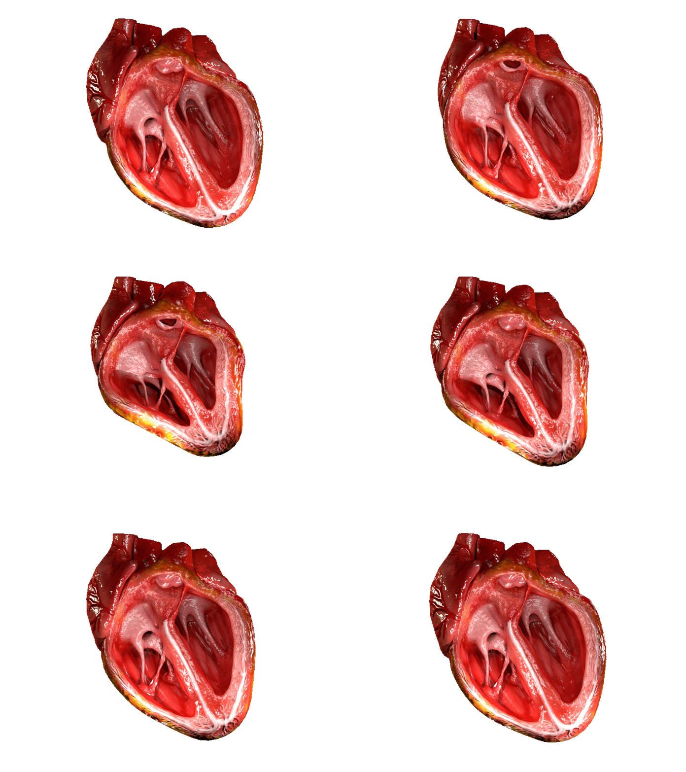
This computer-generated cross-section offers a vivid internal view of a healthy human heart, showcasing its four chambers, robust muscular walls, and the intricate architecture of its valves. This detailed perspective is instrumental in understanding how this vital organ efficiently pumps blood throughout the body. Examining the features of a healthy heart provides a crucial benchmark for identifying deviations that may indicate cardiovascular disease.

The image, depicting a healthy heart in cross-section, allows us to observe several key anatomical features. While specific labels are not present in the image, we can identify and explain the fundamental components based on typical heart anatomy.
Right Atrium: This upper-right chamber is responsible for receiving deoxygenated blood returning from the body through the superior and inferior vena cava. Its relatively thin muscular wall indicates its role as a collecting chamber, pumping blood into the right ventricle.
Left Atrium: Situated in the upper-left, this chamber receives oxygenated blood from the lungs via the pulmonary veins. Like its counterpart, the left atrium primarily functions as a receiving chamber, pushing blood into the left ventricle.
Right Ventricle: This lower-right chamber is characterized by thicker muscular walls than the atria, reflecting its role in pumping deoxygenated blood to the lungs. The presence of the tricuspid valve, controlling flow from the right atrium, and the pulmonary valve, controlling outflow to the pulmonary artery, is implied.
Left Ventricle: As the most muscular and largest chamber, the left ventricle is prominently displayed, possessing significantly thicker walls than any other chamber. Its powerful contractions are responsible for ejecting oxygenated blood into the aorta, propelling it throughout the entire systemic circulation.
Tricuspid Valve: Located between the right atrium and the right ventricle, this valve is crucial for preventing the backflow of blood into the right atrium during ventricular contraction. Its structure, typically with three leaflets, is implied by the visible opening and chordae tendineae.
Mitral Valve (Bicuspid Valve): Positioned between the left atrium and the left ventricle, this valve ensures unidirectional flow of oxygenated blood. Its two leaflets, along with their associated chordae tendineae and papillary muscles, are critical for preventing backflow into the left atrium during the left ventricular systole.
Interventricular Septum: This thick muscular wall separates the right and left ventricles, preventing the mixing of deoxygenated and oxygenated blood. Its integrity is fundamental for the efficient functioning of the heart as a double pump.
The human heart is a remarkable muscular organ, functioning as a dual pump to ensure the continuous circulation of blood. This sophisticated 3D cross-section beautifully illustrates its four distinct chambers, separated by an interventricular septum and atrioventricular valves. The right side of the heart manages deoxygenated blood from the body, directing it towards the lungs for oxygenation, while the left side receives oxygenated blood from the lungs and propels it to every other part of the body. This strict separation and precise coordination of chambers and valves are hallmarks of a healthy cardiovascular system.
The efficiency of this pumping action relies heavily on the structural integrity and synchronized operation of the heart’s valves. Each valve acts as a one-way gate, opening and closing in perfect rhythm to prevent blood from flowing backward. For instance, the robust construction of the left ventricle and its associated mitral and aortic valves are critical for generating the high pressure required for systemic circulation. The visual clarity of these internal structures provides invaluable insight into the mechanics of blood flow, enabling a deeper understanding of cardiac physiology.
Understanding the anatomy of a healthy heart is the foundational step in identifying and diagnosing various cardiovascular diseases. Conditions like valvular stenosis (narrowing) or regurgitation (leakage) directly impede the efficient flow of blood, placing undue stress on the heart. For example, if the mitral valve does not close completely, blood can leak back into the left atrium, increasing atrial pressure and potentially leading to heart failure over time. Therefore, recognizing the normal structure helps in pinpointing pathological changes.
- The heart beats approximately 100,000 times a day in an adult.
- The muscular walls of the left ventricle are typically three times thicker than those of the right ventricle.
- The “lub-dub” sounds of the heartbeat are caused by the closing of the heart valves.
- The human heart can pump about 5 liters of blood per minute at rest.
This detailed 3D rendering of a healthy heart’s cross-section serves as an invaluable educational tool for both medical students and the general public. It underscores the incredible complexity and precision required for this vital organ to sustain life. A thorough appreciation of its normal internal structures and functions is paramount for promoting cardiovascular health and recognizing the importance of early detection and treatment of heart conditions.

