Delve into the intricate internal workings of the human heart with this detailed anatomical view, showcasing its chambers, valves, and major blood vessels. This exploration will illuminate how this vital organ efficiently pumps blood throughout the body, a process fundamental to sustaining life. Understanding the heart’s internal architecture is key to comprehending its function and the origins of various cardiovascular conditions.
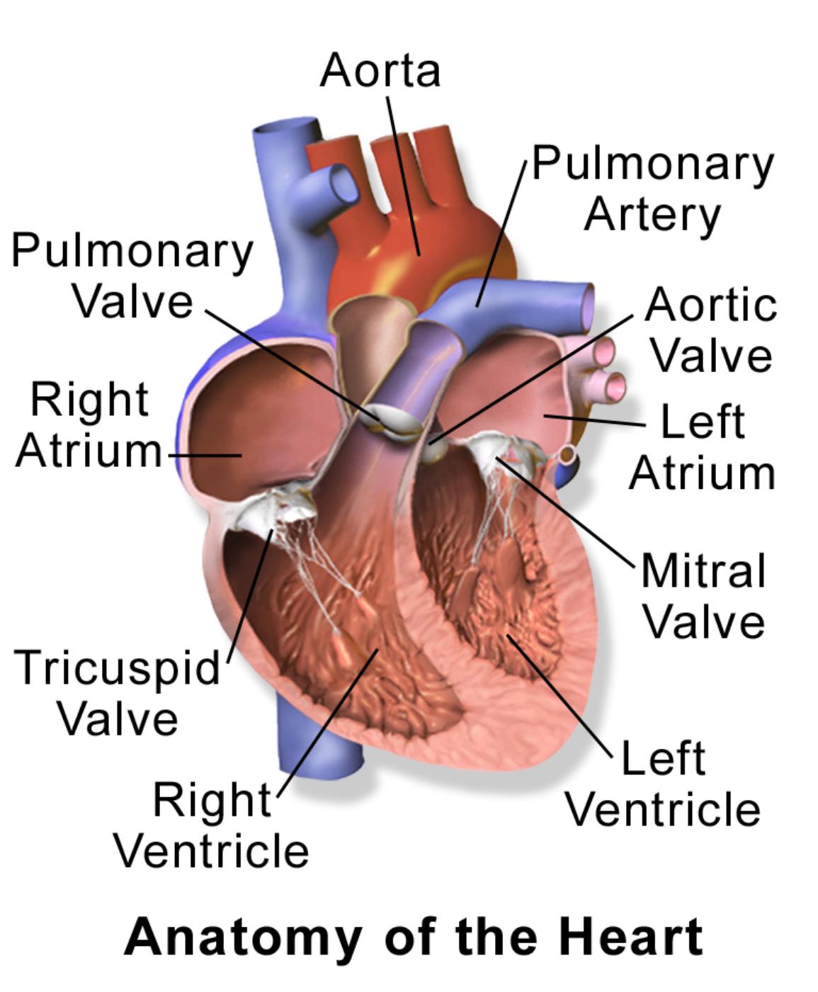
Aorta: The aorta is the largest artery in the body, originating from the left ventricle of the heart and extending down to the abdomen. It is responsible for distributing oxygenated blood to all parts of the body through systemic circulation.
Pulmonary Artery: The pulmonary artery carries deoxygenated blood from the right ventricle of the heart to the lungs. It is unique among arteries as it carries deoxygenated blood.
Pulmonary Valve: The pulmonary valve is one of the four heart valves, located between the right ventricle and the pulmonary artery. It opens to allow blood to flow from the right ventricle to the pulmonary artery and closes to prevent backflow into the right ventricle.
Aortic Valve: The aortic valve is situated between the left ventricle and the aorta, regulating the flow of oxygenated blood out of the heart. Its proper functioning is critical for maintaining systemic blood pressure and circulation.
Right Atrium: The right atrium is the upper right chamber of the heart, receiving deoxygenated blood from the body via the superior and inferior vena cava. It then pumps this blood into the right ventricle.
Left Atrium: The left atrium is the upper left chamber of the heart, receiving oxygenated blood from the lungs via the pulmonary veins. It then pumps this oxygen-rich blood into the left ventricle.
Mitral Valve: Also known as the bicuspid valve, the mitral valve is located between the left atrium and the left ventricle. It ensures that oxygenated blood flows in one direction from the atrium to the ventricle, preventing backflow during ventricular contraction.
Tricuspid Valve: The tricuspid valve is situated between the right atrium and the right ventricle. It prevents the backflow of deoxygenated blood into the right atrium when the right ventricle contracts.
Right Ventricle: The right ventricle is the lower right chamber of the heart, responsible for pumping deoxygenated blood to the lungs through the pulmonary artery. This action is crucial for the process of gas exchange.
Left Ventricle: The left ventricle is the lower left chamber of the heart and is the most muscular chamber, responsible for pumping oxygenated blood into the aorta and to the rest of the body. Its strong contractions generate the high pressure needed for systemic circulation.
The human heart is a remarkable organ, a tireless pump that ensures the continuous circulation of blood, delivering oxygen and nutrients to every cell while removing waste products. This internal view reveals a four-chambered structure, divided into two atria (upper chambers) and two ventricles (lower chambers). This separation is vital for keeping oxygenated and deoxygenated blood from mixing, a crucial aspect of efficient circulation. The rhythmic contraction and relaxation of these chambers, orchestrated by a complex electrical system, propel blood through a vast network of vessels.
Blood flow through the heart is a meticulously regulated process, guided by a system of four valves that act as one-way gates. These valves open and close in precise coordination, preventing the backflow of blood and ensuring its unidirectional movement. The pulmonary and aortic valves control blood flow out of the ventricles, while the tricuspid and mitral valves regulate flow between the atria and ventricles. The integrity and proper functioning of these valves are paramount for maintaining cardiovascular health.
Any compromise to these valves, such as stenosis (narrowing) or regurgitation (leakage), can significantly impact the heart’s efficiency and lead to various cardiovascular diseases. For example, mitral valve regurgitation, where the mitral valve does not close completely, allows blood to leak back into the left atrium, increasing the workload on the heart. Understanding the precise location and function of each valve is critical for diagnosing and treating such conditions.
- The heart beats approximately 100,000 times a day.
- The total length of blood vessels in an adult human is about 60,000 miles.
- Heart valves open and close due to pressure differences.
- The left ventricle generates the highest pressure in the heart.
This internal anatomical perspective not only provides a foundational understanding of cardiac physiology but also underscores the delicate balance required for optimal heart function. From the reception of blood in the atria to its powerful ejection from the ventricles, every component plays a pivotal role in sustaining life. A comprehensive grasp of these internal structures is indispensable for both medical education and for individuals seeking a deeper insight into their own health.
Understanding the intricate internal anatomy of the heart, with its specialized chambers, valves, and major blood vessels, is fundamental to appreciating its profound role in human physiology. This detailed view allows us to grasp the elegance and efficiency with which this vital organ operates, ensuring the constant circulation of life-sustaining blood. The coordinated efforts of each component highlight the importance of maintaining cardiovascular health for overall well-being.

