Delve into the intricate world of early avian embryology through a transverse section of a 45-hour chick embryo, revealing the foundational tissue layers and developing structures. This article explores the formation of critical organs and systems, offering insights into the complex processes that shape a vertebrate organism. Understand how the ectoderm, mesoderm, and endoderm orchestrate the emergence of neural pathways, somites, and the primitive coelom.
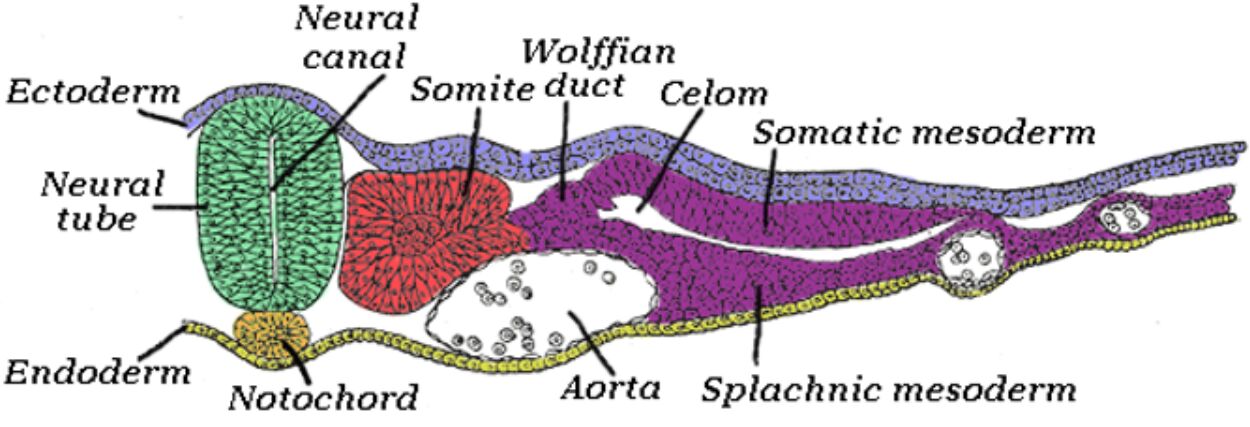
Ectoderm: This is the outermost of the three primary germ layers in the early embryo, responsible for forming the epidermis (skin), hair, feathers, nervous system, and sensory organs. In this transverse section, it is visible as the outermost layer, contributing to the initial formation of the neural tube.
Neural tube: A hollow structure formed from the folding of the neural plate (derived from the ectoderm), the neural tube is the embryonic precursor to the central nervous system, including the brain and spinal cord. Its formation, a process called neurulation, is a critical step in vertebrate development, indicating the future axis of the nervous system.
Neural canal: This is the central lumen or cavity within the neural tube, which will eventually develop into the ventricles of the brain and the central canal of the spinal cord. It is filled with cerebrospinal fluid and plays a crucial role in the circulation of nutrients and waste products within the central nervous system.
Endoderm: The innermost of the three primary germ layers, the endoderm gives rise to the lining of the digestive tract and its associated glands (like the liver and pancreas), as well as the respiratory system. In the image, it forms the ventral-most layer, underlying the notochord.
Notochord: A flexible, rod-like structure derived from the mesoderm, the notochord serves as the primary axial support in early vertebrate embryos and induces the formation of the neural tube. While largely replaced by the vertebral column in most adult vertebrates, it persists in remnants and is vital for embryonic signaling.
Somite duct (also known as pronephric duct or nephric duct): This embryonic excretory duct, forming from the intermediate mesoderm, is a precursor to parts of the kidney and urinary system. It plays a role in early waste removal and development of the excretory system in vertebrates.
Wolffian (Mesonephric) duct: This structure, developing from the pronephric duct, is a crucial part of the embryonic urinary and male reproductive systems in many vertebrates. It contributes to the formation of the mesonephros (a temporary kidney) and subsequently forms the vas deferens in males.
Celom: This refers to the main body cavity that develops within the mesoderm, eventually housing and protecting various internal organs such as the heart, lungs, and digestive organs. Its formation is a significant step in the organization of the vertebrate body plan, allowing for organ system development and movement.
Somatic mesoderm: This is the outer layer of the lateral plate mesoderm, located adjacent to the ectoderm. It contributes to the formation of the body wall, limbs, and the parietal peritoneum (lining of the coelomic cavity).
Aorta: In an embryonic context, the aorta represents the primitive dorsal aorta, which is the main arterial trunk supplying oxygenated blood to the developing tissues. It signifies the nascent cardiovascular system, essential for nutrient and gas exchange.
Splanchnic mesoderm: This is the inner layer of the lateral plate mesoderm, located adjacent to the endoderm. It gives rise to the visceral peritoneum (lining of the organs within the coelom), the smooth muscle and connective tissue of the digestive tract, and the heart.
The development of a vertebrate embryo is a meticulously orchestrated process, transforming a single fertilized cell into a complex multicellular organism. The chick embryo, a classic model in developmental biology, provides an accessible window into these fundamental stages. By 45 hours of incubation, the chick embryo has undergone significant morphological changes, establishing the basic body plan and initiating the formation of crucial organ systems. A transverse section at this stage reveals the primary germ layers—ectoderm, mesoderm, and endoderm—and their intricate interactions, laying down the groundwork for future tissues and organs.
At this developmental juncture, the central nervous system is rapidly progressing, evident in the prominent neural tube derived from the ectoderm. This structure, which will ultimately give rise to the brain and spinal cord, encloses the neural canal, a precursor to the ventricular system. Concurrently, the mesoderm, positioned between the ectoderm and endoderm, is undergoing differentiation into various sub-regions. The somites, segmental blocks of mesoderm, are particularly visible, signaling the future development of vertebrae, muscles, and dermis. These structures highlight the axial organization that is characteristic of vertebrate embryos.
The formation of the coelom, the primary body cavity, from the lateral plate mesoderm is also a critical event by 45 hours. This cavity provides space for organ development and movement within the body. Simultaneously, the early circulatory system, represented by the primitive aorta, is beginning to establish, ensuring the transport of nutrients and oxygen to the rapidly growing tissues. The interplay between these developing structures—from the notochord providing axial support and inducing neural tube formation, to the differentiation of the mesoderm into components like the Wolffianduct—underscores the complexity and interdependence of embryonic processes.
Key developmental structures visible at 45 hours include:
- Neural tube and canal: Forming the CNS.
- Notochord: Axial support and inductive signaling.
- Somites: Precursors to axial skeleton, muscles, dermis.
- Coelom: Body cavity formation.
- Primitive aorta: Early circulatory system.
- Wolffian (pronephric) duct: Early excretory system.
In conclusion, the transverse section of a 45-hour chick embryo offers a remarkable snapshot of early vertebrate development, showcasing the precise coordination of cellular and tissue differentiation. The clear delineation of the ectoderm, mesoderm, and endoderm, alongside the formation of the neural tube, notochord, somites, and nascent circulatory and excretory systems, illustrates the fundamental anatomical blueprint being established. This intricate dance of embryonic processes highlights the elegance and complexity inherent in the genesis of a complete organism, providing invaluable insights into developmental biology and the continuity of life.
ule.

