Unravel the anatomical mystery of how both the giraffe and its shorter-necked cousin, the okapi, possess precisely seven cervical vertebrae, despite their dramatic differences in neck length. This article explores the fascinating role of heterochrony in shaping these iconic mammalian necks, providing insights into evolutionary development and vertebrate anatomy.
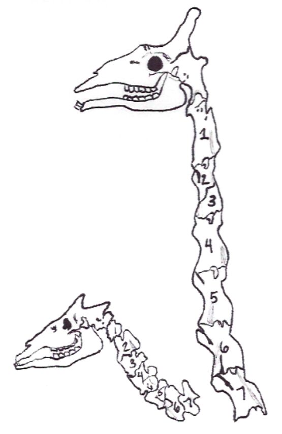
Okapi (left) and giraffe (right): These labels distinguish between the two animals depicted, illustrating their distinct neck lengths while highlighting their shared fundamental cervical vertebral count. The okapi, a forest-dwelling ruminant, possesses a neck length more typical of other mammals, whereas the giraffe is renowned for its extraordinarily elongated neck.
Seven cervical vertebrae structure: This refers to the consistent number of bones (C1-C7) forming the neck region in both the okapi and the giraffe, despite the immense difference in their overall neck dimensions. This numerical constancy is a remarkable example of developmental constraint within mammalian evolution, where the length is achieved through individual vertebral elongation rather than an increase in count.
The mammalian neck is a region of immense anatomical variation, yet a striking consistency often observed across species is the number of cervical vertebrae. From a mouse to a whale, and as vividly illustrated by the giraffe and its close relative, the okapi, the vast majority of mammals possess precisely seven cervical vertebrae. This conserved number, irrespective of the dramatic differences in neck length, presents a compelling case study in evolutionary development and the mechanisms that shape animal forms. The image juxtaposes the short, muscular neck of the okapi with the famously elongated neck of the giraffe, both revealing this fundamental anatomical blueprint.
The profound difference in neck length between the giraffe and the okapi, despite sharing the same number of cervical vertebrae, is primarily attributed to a developmental phenomenon known as heterochrony. Heterochrony refers to a change in the timing or rate of developmental events during an organism’s ontogeny relative to its ancestors or other closely related species. In the case of the giraffe, the extreme elongation of its neck bones (specifically the individual cervical vertebrae) is a result of an extended period of embryonic development for these particular skeletal elements. This extended growth phase allows each of the seven cervical vertebrae to reach a far greater length compared to those of the okapi or other mammals.
This evolutionary adaptation in giraffes has conferred significant ecological advantages, primarily enabling them to browse on foliage inaccessible to other herbivores. The elongated neck, while advantageous, also presents unique physiological challenges, such as the need for a powerful cardiovascular system to pump blood to the brain and specialized vascular controls to prevent blackouts when lowering the head. The okapi, conversely, has maintained a more ancestral neck length, suited to its browsing habits in dense forest undergrowth. Both species, however, perfectly demonstrate how evolutionary pressures can manipulate developmental pathways to produce remarkable phenotypic diversity from a shared anatomical foundation.
Key aspects of cervical vertebrae in these animals include:
- Constant count: Always seven vertebrae (C1-C7).
- Variable length: Individual vertebrae greatly elongated in giraffes.
- Heterochrony: Developmental mechanism for elongation.
- Functional adaptation: Browsing high foliage (giraffe) vs. dense undergrowth (okapi).
- Physiological implications: Blood pressure regulation in giraffes.
In conclusion, the comparative anatomy of the okapi and giraffe cervical spines beautifully illustrates a fundamental principle of mammalian evolution: developmental processes can be subtly altered to produce dramatically different phenotypes from a conserved genetic and anatomical blueprint. The consistent presence of seven cervical vertebrae, coupled with the profound impact of heterochrony on their individual growth, highlights the elegant solutions evolution devises to adapt species to their unique ecological niches. This understanding is crucial for appreciating the intricate interplay between development, anatomy, and adaptation in the natural world.

