Unraveling the Goat’s Backbone: A Comprehensive Guide to Vertebral Anatomy
Discover the intricate design of the goat’s vertebral column, a masterpiece of natural engineering that underpins its agility and strength. This article delves into the specific regions of a goat’s vertebrae, offering a detailed look at how each section contributes to the animal’s overall movement and support. Understanding these anatomical distinctions is key to appreciating the biomechanics of these remarkable ruminants.
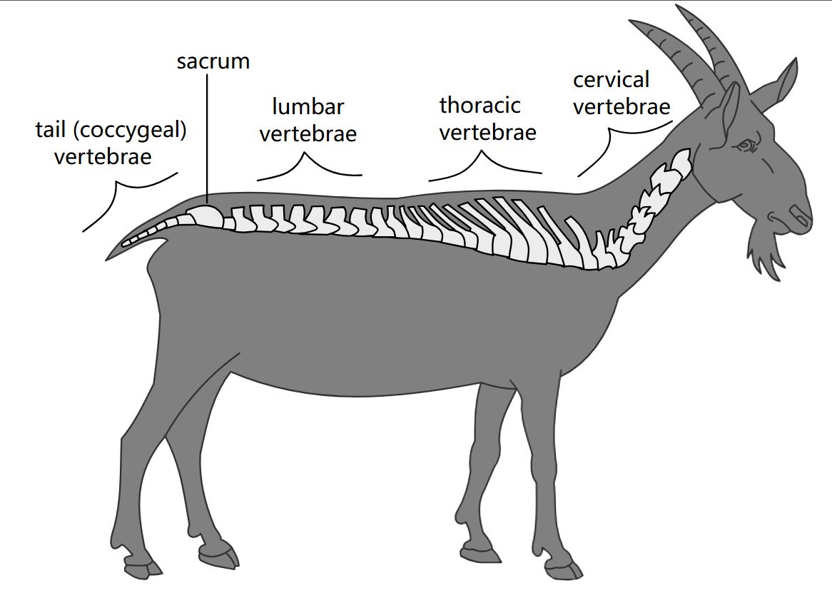
Cervical Vertebrae: The cervical vertebrae form the neck region of the goat, playing a crucial role in head movement and flexibility. These vertebrae are typically larger and more robust, designed to support the head and allow for a wide range of motion, essential for grazing and alertness.
Thoracic Vertebrae: Located in the chest area, the thoracic vertebrae are characterized by their longer spinous processes, which connect to the ribs. This section of the spine provides a sturdy framework for the rib cage, protecting vital organs and contributing to the animal’s respiratory mechanics.
Lumbar Vertebrae: The lumbar vertebrae constitute the lower back region, offering significant support and flexibility for the goat’s powerful hindquarters. These vertebrae are typically larger than the thoracic vertebrae, designed to withstand considerable stress during locomotion and jumping.
Sacrum: The sacrum is a fused triangular bone located at the base of the spine, connecting the vertebral column to the pelvic girdle. This robust structure is vital for transmitting weight from the body to the hind limbs, providing stability and facilitating powerful movements.
Tail (Coccygeal) Vertebrae: The tail (coccygeal) vertebrae form the caudal appendage of the goat, varying in number and size among individuals. While offering less structural support than other regions, these vertebrae allow for tail movement, which can be used for balance and communication.
The vertebral column, often referred to as the backbone or spine, is a defining characteristic of vertebrates, including the domestic goat (Capra hircus). This complex anatomical structure is not merely a stack of bones but a highly integrated system providing the primary axial support for the body. It encases and protects the delicate spinal cord, which is a vital conduit for nerve signals between the brain and the rest of the body. Furthermore, the spine serves as a crucial attachment point for numerous muscles and ligaments, facilitating movement, maintaining posture, and absorbing shock during various physical activities.
Understanding the specific regions of the goat’s vertebral column offers profound insights into its unique physiology and adaptive capabilities. Each segment of the spine has evolved distinct morphological features tailored to its functional demands. For instance, the neck vertebrae are designed for maximal flexibility, allowing the goat to navigate varied terrain and access forage, while the robust lumbar region supports the powerful muscles essential for climbing and jumping. The intricate interplay between bone, cartilage, and soft tissues within the spine ensures both rigidity and elasticity, enabling the goat to exhibit its characteristic agility and resilience in diverse environments.
The architectural design of the goat’s vertebral column exemplifies the principles of biomechanical efficiency. From the atlas and axis in the cervical region, which facilitate head rotation, to the fused sacral vertebrae that integrate with the pelvis, every component plays a specific role. This segmental organization allows for localized movement without compromising the overall structural integrity of the spine. The intervertebral discs situated between adjacent vertebrae act as natural shock absorbers, protecting the spinal column from impact forces and ensuring smooth, fluid motion. The health and integrity of these vertebral regions are paramount to the goat’s overall well-being and athletic performance.
The goat’s vertebral column can be divided into distinct regions:
- Cervical Region: The neck, highly flexible.
- Thoracic Region: The chest, connected to the ribs.
- Lumbar Region: The lower back, robust and supportive.
- Sacral Region: Fused bones connecting to the pelvis.
- Coccygeal Region: The tail, varying in length.
In conclusion, the detailed examination of the goat’s vertebral column, from the flexible cervical vertebrae to the supportive sacrum and the articulate tail, reveals a marvel of anatomical adaptation. Each region is uniquely specialized, yet all work in concert to provide the structural integrity, protective enclosure for the spinal cord, and mobility essential for the goat’s survival and characteristic behaviors. This understanding not only deepens our appreciation for animal physiology but also provides a foundational knowledge for veterinary science and animal husbandry practices, ensuring the health and welfare of these important livestock animals.

