The formation of urine is a dynamic and highly regulated process within the kidney’s functional unit, the nephron, involving continuous secretion and reabsorption of various substances. This article provides a comprehensive overview of the locations of secretion and reabsorption in the nephron, illustrating how different segments meticulously fine-tune the composition of filtrate to produce the final urine. Understanding these processes is vital for comprehending fluid and electrolyte balance, waste removal, and the mechanisms underlying kidney diseases.
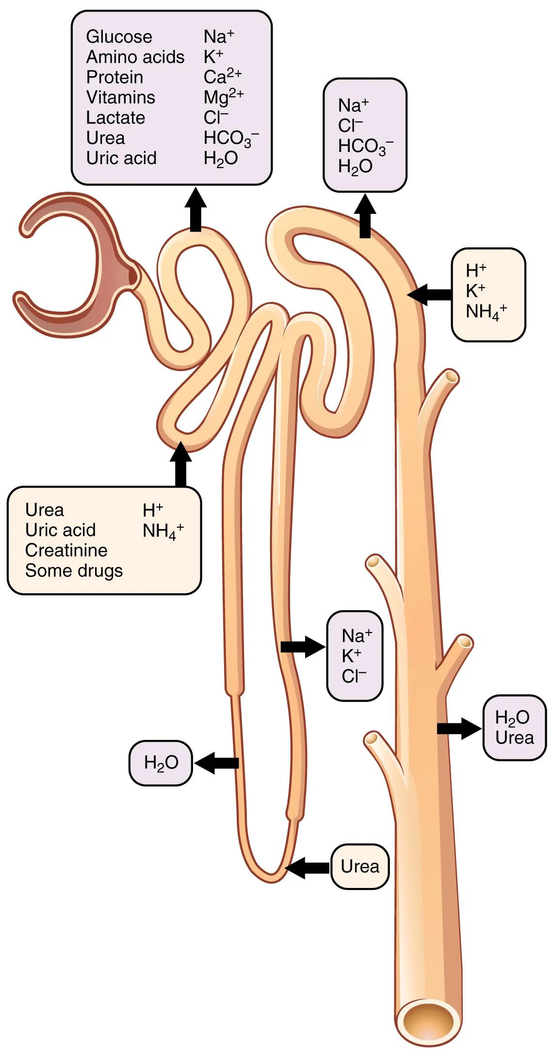
Proximal convoluted tubule (top left box): This initial segment of the renal tubule is a major site for reabsorption. Here, essential nutrients like glucose, amino acids, vitamins, and a significant portion of electrolytes (Na+, K+, Ca2+, Mg2+, Cl-, HCO3-) and water are reclaimed from the filtrate and returned to the bloodstream. Additionally, some waste products such as urea and uric acid are also reabsorbed here.
Distal convoluted tubule / Collecting Duct (top right box): This segment, along with the collecting ducts, is where final adjustments to urine composition occur. Here, reabsorption of Na+, Cl-, HCO3-, and water takes place, often under hormonal control. Simultaneously, substances like H+, K+, and NH4+ are secreted from the blood into the filtrate.
Loop of Henle (middle left box – secretion into tubule): In the descending limb, water is reabsorbed. In the ascending limb, Na+, K+, and Cl- are actively reabsorbed, contributing to the medullary osmotic gradient.
Collecting Duct (bottom right box): The collecting ducts are the final site for water reabsorption, regulated by ADH, and a site for urea reabsorption. They play a critical role in determining the final concentration of urine.
The nephron, the microscopic workhorse of the kidney, is a master of selective filtration, reabsorption, and secretion, ensuring that the body maintains its delicate internal balance. After initial filtration in the glomerulus, the resulting fluid, known as filtrate, embarks on an intricate journey through various segments of the renal tubule. Each segment is equipped with specific transport mechanisms that precisely regulate the movement of water, ions, nutrients, and waste products. This image clearly maps out where these crucial processes of reabsorption (moving substances from filtrate back into blood) and secretion (moving substances from blood into filtrate) predominantly occur, illustrating the dynamic nature of urine formation.
The journey begins in the proximal convoluted tubule (PCT), where the bulk of reabsorption takes place. Here, almost all filtered glucose, amino acids, and vitamins are reabsorbed, along with a significant portion of sodium, potassium, calcium, magnesium, chloride, and bicarbonate ions. Approximately 60-70% of the filtered water is also passively reabsorbed, primarily due to the osmotic pull created by the active transport of solutes. Simultaneously, some waste products like urea and uric acid are partially reabsorbed, while certain organic acids and bases, including some drugs, are actively secreted into the tubule. This segment is essentially a “mass reabsorber,” recovering most of the valuable substances that were initially filtered.
As the filtrate progresses into the loop of Henle, a critical osmotic gradient is established in the renal medulla, enabling the kidney to produce concentrated urine. The descending limb is highly permeable to water, allowing water to exit the tubule and be reabsorbed into the bloodstream. In contrast, the ascending limb is impermeable to water but actively reabsorbs sodium, potassium, and chloride ions. This differential permeability is crucial for establishing the high osmolality of the medullary interstitial fluid. The distal convoluted tubule (DCT) and collecting ducts are the final sites for fine-tuning the filtrate’s composition, where reabsorption and secretion are largely under hormonal control. For example, aldosterone stimulates sodium reabsorption and potassium secretion, while antidiuretic hormone (ADH) regulates water reabsorption by promoting the insertion of aquaporin channels. Here, hydrogen ions and ammonium are also secreted, playing a role in acid-base balance.
Dysfunction in any segment of the nephron’s reabsorption or secretion processes can lead to significant physiological imbalances and renal diseases. For instance, impaired glucose reabsorption in the PCT, often seen in uncontrolled diabetes mellitus, leads to glucose in the urine (glycosuria). Defects in the loop of Henle’s ability to reabsorb salts can impair the kidney’s capacity to concentrate urine, resulting in polyuria (excessive urination). Furthermore, imbalances in the hormonal control of the DCT and collecting ducts, such as in cases of inadequate ADH production (diabetes insipidus), can lead to severe dehydration. Understanding these precise locations and mechanisms of reabsorption and secretion is therefore indispensable for diagnosing, treating, and managing a wide array of renal pathologies, ensuring the kidney can effectively maintain the body’s fluid, electrolyte, and acid-base homeostasis.

