Fenestrated capillaries are a specialized type of blood vessel crucial for rapid fluid and solute exchange in specific organs. This article explores the unique fenestrated capillary anatomical structure, highlighting the presence of pores that facilitate efficient filtration. Understanding these capillaries is essential for grasping their vital roles in kidney function, endocrine glands, and other sites requiring rapid transport of substances.
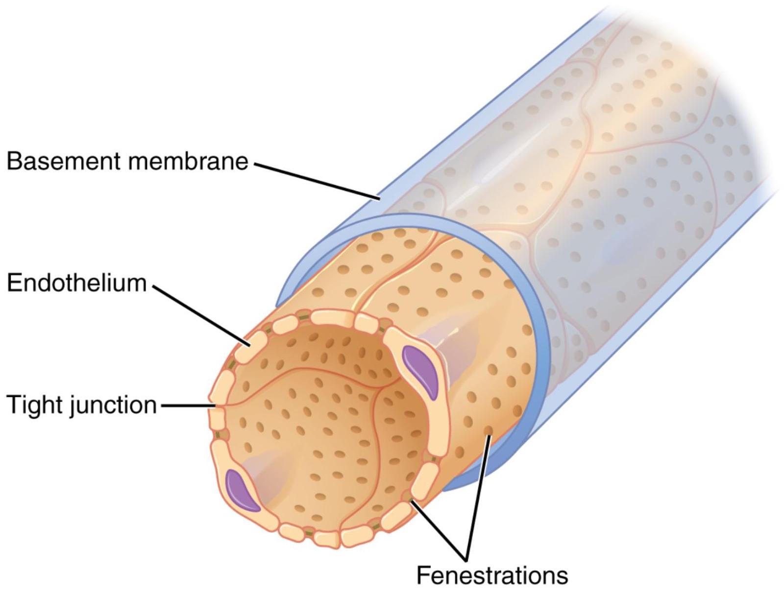
Basement membrane: This is a thin, extracellular layer that provides structural support to the endothelium and acts as a selective barrier. It is composed of collagen and glycoproteins, playing a critical role in filtering substances based on size and charge.
Endothelium: This is the innermost layer of cells lining the capillary lumen, forming the primary barrier between blood and surrounding tissues. In fenestrated capillaries, these endothelial cells are characterized by the presence of numerous pores.
Tight junction: These are specialized intercellular junctions that form a watertight seal between adjacent endothelial cells. While some capillaries have them, in fenestrated capillaries, the presence of fenestrations allows for rapid passage of substances even if the tight junctions are present to maintain cell-to-cell adhesion.
Fenestrations: These are small, open pores or windows within the endothelial cells of the capillary wall. They significantly increase the permeability of the capillary, allowing for the rapid diffusion of water and small solutes from the blood.
Capillaries are the smallest blood vessels, serving as the primary sites for the exchange of nutrients, waste products, and gases between blood and tissues. While all capillaries are designed for exchange, not all are created equal. Fenestrated capillaries represent a specialized subtype, uniquely adapted for situations requiring exceptionally rapid and efficient filtration or absorption. These capillaries are strategically located in organs where such rapid exchange is paramount, underscoring their critical role in maintaining various physiological processes. The image clearly depicts the distinctive features of these vessels, particularly the presence of pores within their endothelial lining.
The defining characteristic of fenestrated capillaries is the presence of numerous pores, or fenestrations, within the endothelial cells that form their walls. These fenestrations typically range from 60-80 nanometers in diameter and are often covered by a thin, permeable diaphragm, except in the renal glomeruli. This structural adaptation significantly increases the permeability of the capillary wall compared to continuous capillaries, allowing for the quick passage of water and small solutes. Surrounding the endothelium is a basement membrane, a non-cellular layer that acts as an additional selective filter, restricting the movement of larger molecules. This two-part filtration system ensures that while rapid exchange occurs, certain essential components of the blood, such as large proteins and blood cells, are retained.
Fenestrated capillaries are found in several key locations throughout the body, each highlighting their specialized function:
- Kidneys (Glomeruli): Here, they are crucial for the initial formation of urine. The fenestrations allow large volumes of plasma to be filtered from the blood into Bowman’s capsule, while the basement membrane and podocytes prevent the passage of proteins.
- Endocrine Glands: In glands like the thyroid, pituitary, and adrenal glands, fenestrated capillaries facilitate the rapid uptake and release of hormones into the bloodstream.
- Intestinal Villi: In the small intestine, they play a role in the absorption of digested nutrients from the lumen into the blood.
Damage or dysfunction of fenestrated capillaries can have significant implications for health. In the kidneys, for example, conditions that affect the integrity of the glomerular capillaries and their fenestrations can lead to impaired filtration, proteinuria (presence of excess protein in urine), and ultimately kidney disease. Diabetic nephropathy, a common complication of diabetes, involves thickening of the glomerular basement membrane and changes to the fenestrated endothelium, leading to reduced filtration efficiency. Understanding the precise structure and function of fenestrated capillaries is therefore vital for diagnosing and managing conditions that impact fluid balance, hormone regulation, and waste elimination, underscoring their fundamental role in systemic health.

