The nephron is the fundamental functional unit of the kidney, a microscopic marvel responsible for filtering blood, reabsorbing essential substances, and ultimately forming urine. This article delves into the intricate process of blood flow in the nephron, highlighting how each specialized segment contributes to maintaining the body’s delicate internal balance. Understanding the precise interplay between blood vessels and renal tubules is key to comprehending kidney function and the mechanisms behind renal health and disease.
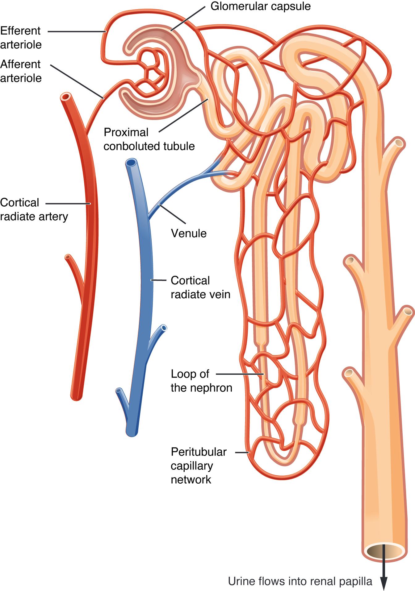
Efferent arteriole: This small artery carries blood away from the glomerulus, after the initial filtration process has occurred. It is narrower than the afferent arteriole, which helps maintain high pressure in the glomerulus.
Afferent arteriole: This small artery branches from a cortical radiate artery and carries unfiltered blood into the glomerulus. Its diameter is larger than the efferent arteriole, contributing to the pressure required for filtration.
Glomerular capsule (also known as Bowman’s capsule): This cup-shaped structure surrounds the glomerulus and collects the filtrate produced during blood filtration. It is the beginning of the tubular component of the nephron.
Proximal convoluted tubule: This is the first segment of the renal tubule, extending from the glomerular capsule. It is responsible for the reabsorption of a large percentage of filtered water, ions, organic nutrients, and proteins back into the blood.
Cortical radiate artery: These small arteries branch off the arcuate arteries and extend into the renal cortex, supplying blood to the nephrons. They give rise to the afferent arterioles.
Venule: These small veins collect deoxygenated blood from the peritubular capillaries and eventually drain into larger veins. They are part of the venous drainage system of the kidney.
Cortical radiate vein: These small veins collect blood from the venules in the renal cortex. They ultimately drain into the arcuate veins.
Loop of the nephron (also known as the loop of Henle): This U-shaped segment of the renal tubule extends into the renal medulla, playing a crucial role in creating the osmotic gradient necessary for concentrating urine. It has descending and ascending limbs with differing permeabilities to water and solutes.
Peritubular capillary network: This extensive network of capillaries surrounds the renal tubules, particularly in the cortex. It is vital for the reabsorption of water and solutes from the filtrate back into the blood, and for the secretion of waste products into the tubules.
The human body relies on the kidneys to filter approximately 180 liters of blood plasma daily, a monumental task performed by millions of microscopic units called nephrons. Each nephron is a sophisticated mini-filter and processing plant, ensuring that waste products are eliminated while essential substances are conserved. This intricate process is initiated by a unique vascular arrangement that facilitates the initial filtration of blood, followed by a tubular system designed for precise reabsorption and secretion. The image vividly illustrates the close anatomical and functional relationship between the blood vessels and the renal tubules within a single nephron, highlighting the efficiency of this vital organ.
The journey of blood through the nephron begins with the afferent arteriole delivering unfiltered blood to the glomerulus, a specialized capillary bed encased within the glomerular capsule. Here, due to the high hydrostatic pressure, a process called glomerular filtration occurs, where water and small solutes are forced out of the blood and into the capsule, forming the initial filtrate. This filtrate is essentially plasma without large proteins and blood cells. The unique diameter difference between the afferent and efferent arterioles helps maintain this crucial filtration pressure. The efferent arteriole then carries the remaining blood, now concentrated with proteins and cells, away from the glomerulus.
Following filtration, the filtrate enters the renal tubule, where its composition undergoes significant modification. The proximal convoluted tubule is the primary site for the reabsorption of most filtered substances, including glucose, amino acids, and a large proportion of water and ions, back into the bloodstream via the peritubular capillaries. As the filtrate moves into the loop of the nephron, a critical osmotic gradient is established in the renal medulla, which is essential for concentrating the urine. The ascending limb actively transports salts out of the tubule, while the descending limb is permeable to water, allowing for further water reabsorption. Finally, in the distal convoluted tubule and collecting duct, fine-tuning of water and electrolyte balance occurs, influenced by hormones like ADH and aldosterone, before the refined urine flows into the renal papilla.
Disruptions to the delicate balance of blood flow and tubular function within the nephron can lead to a spectrum of renal diseases. For instance, hypertension can damage the afferent and efferent arterioles and the glomerulus itself, impairing filtration and leading to chronic kidney disease. Diabetes mellitus, if poorly controlled, can cause glomerulosclerosis, a scarring of the glomeruli, further compromising their filtering capacity. Conditions affecting the peritubular capillaries, such as vasculitis, can disrupt the reabsorption and secretion processes. Understanding the precise mechanisms of blood flow within the nephron and its functional segments is therefore paramount for accurate diagnosis, effective treatment, and ultimately, the prevention of progressive kidney damage, underscoring the vital role of this microscopic structure in systemic health.

