Explore the intricate neural network within the brainstem that precisely controls our breathing, from quiet respiration to forced movements. This article delves into the roles of the medulla and pons, highlighting the specific respiratory groups and centers that coordinate the diaphragm and intercostal muscles for life-sustaining air exchange.
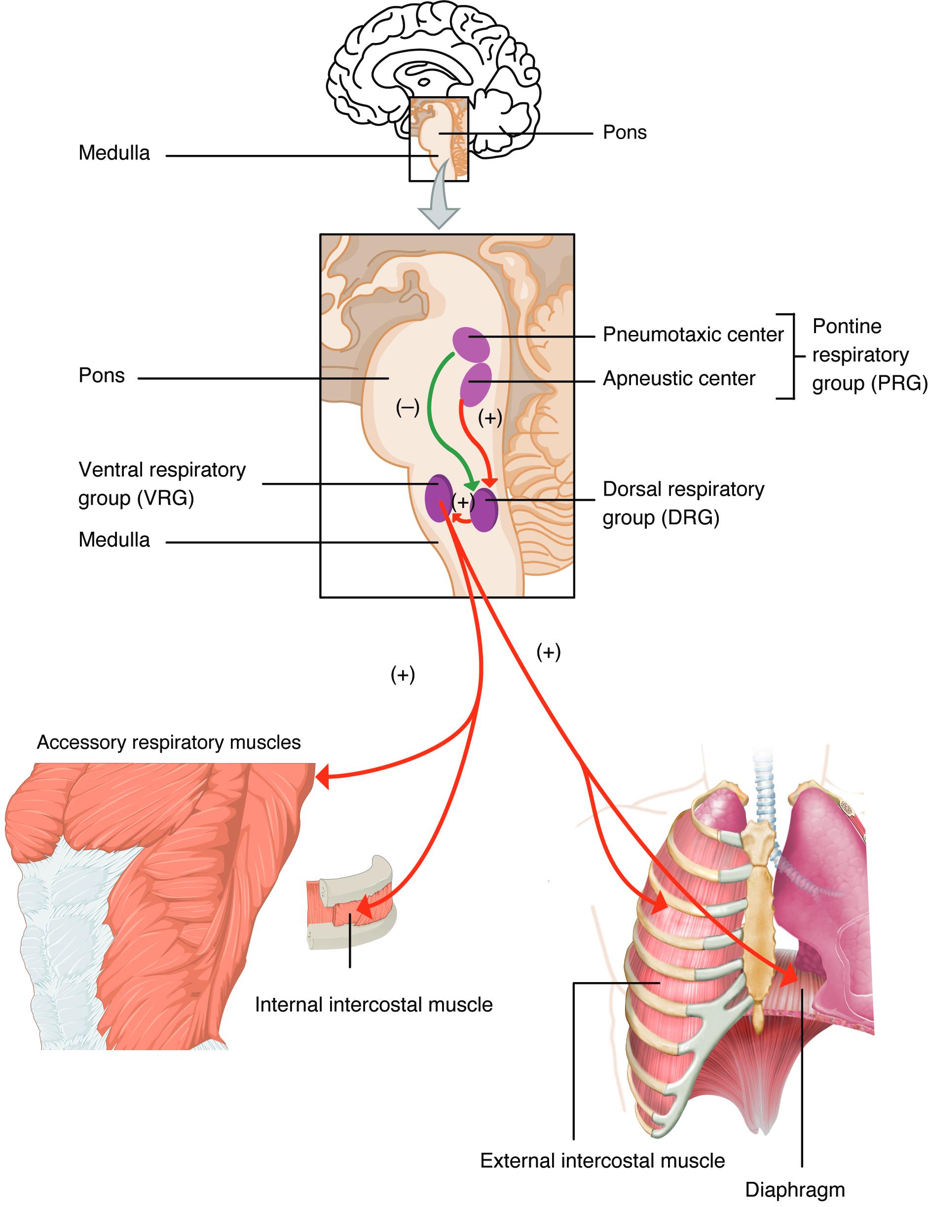
Medulla: The medulla oblongata is a crucial part of the brainstem responsible for regulating several fundamental autonomic functions, including breathing, heart rate, and blood pressure. It contains the primary respiratory rhythm-generating centers, specifically the dorsal and ventral respiratory groups.
Pons: The pons is another critical component of the brainstem, situated superior to the medulla. It contains the pontine respiratory group, which includes the pneumotaxic and apneustic centers, both influencing the rate and depth of breathing.
Pneumotaxic center: Located in the upper pons, this center primarily sends inhibitory signals to the inspiratory neurons in the medulla. Its main function is to limit inspiration, thereby increasing the respiratory rate and promoting shorter, shallower breaths.
Apneustic center: Situated in the lower pons, this center provides stimulatory impulses to the inspiratory neurons of the dorsal respiratory group in the medulla. It promotes prolonged inspiration, leading to deep and sustained breaths, a condition known as apneusis if unregulated.
Pontine respiratory group (PRG): This group, located in the pons, consists of the pneumotaxic and apneustic centers. It modifies the activity of the medullary respiratory centers, ensuring a smooth and coordinated breathing pattern.
Ventral respiratory group (VRG): Located in the medulla, the VRG contains both inspiratory and expiratory neurons. While relatively inactive during quiet breathing, it becomes crucial during forced breathing, sending signals to accessory respiratory muscles for powerful inhalation and exhalation.
Dorsal respiratory group (DRG): Also found in the medulla, the DRG is the primary rhythm-generating center for respiration. It mainly contains inspiratory neurons that send signals to the diaphragm and external intercostal muscles, initiating normal, quiet inspiration.
Accessory respiratory muscles: These muscles, located in the neck and chest, are not primarily involved in quiet breathing but are recruited during forced or strenuous respiration. Examples include the sternocleidomastoid and scalene muscles for inspiration, and abdominal muscles for forced expiration.
Internal intercostal muscle: Located between the ribs, these muscles are primarily involved in forced expiration. When they contract, they pull the ribs inward and downward, helping to rapidly decrease the volume of the thoracic cavity.
External intercostal muscle: Situated between the ribs, these muscles are primarily responsible for inspiration. Their contraction pulls the ribs upward and outward, expanding the thoracic cavity and drawing air into the lungs during normal and forced inhalation.
Diaphragm: The diaphragm is the most important muscle of respiration, a large, dome-shaped muscle separating the thoracic and abdominal cavities. Its contraction flattens it, increasing thoracic volume and initiating inspiration.
The Neurological Symphony of Breathing: An Overview
Respiration, a fundamental physiological process, is meticulously controlled by an intricate network of specialized nerve cells located within the brainstem. This neural command center ensures that breathing is both rhythmic and adaptable, capable of adjusting to the body’s varying demands for oxygen and carbon dioxide expulsion. From the unconscious breaths we take while sleeping to the rapid, deep inhalations during strenuous exercise, the respiratory centers of the brain work tirelessly to maintain vital gas exchange.
The primary regions involved in orchestrating this complex process are the medulla oblongata and the pons. These two components of the brainstem house distinct groups of neurons that generate the basic breathing rhythm and fine-tune its pattern. Understanding the functions of these centers is crucial for appreciating how our body maintains oxygen homeostasis and responds to changes in metabolic activity or environmental conditions.
The brain’s respiratory control involves:
- Generating the fundamental rhythm of breathing.
- Modulating the rate and depth of respiration.
- Coordinating the activity of respiratory muscles.
This sophisticated control system ensures that breathing is a seamlessly integrated process, vital for sustaining life.
The Medulla: The Heartbeat of Respiration
The medulla oblongata is the primary site for generating the basic rhythm of breathing. It contains two crucial collections of neurons: the Dorsal Respiratory Group (DRG) and the Ventral Respiratory Group (VRG).
Dorsal Respiratory Group (DRG)
The Dorsal Respiratory Group (DRG), located in the dorsal portion of the medulla, is the fundamental rhythm generator for normal, quiet breathing. It primarily consists of inspiratory neurons that spontaneously fire, sending excitatory signals to the diaphragm and external intercostal muscles. These signals cause the muscles to contract, leading to inspiration. When the DRG neurons cease firing, these muscles relax, and passive expiration occurs. The DRG receives input from various sources, including chemoreceptors (detecting changes in blood gas levels like oxygen and carbon dioxide) and mechanoreceptors (detecting lung stretch), allowing it to modify the breathing pattern in response to physiological needs.
Ventral Respiratory Group (VRG)
The Ventral Respiratory Group (VRG), situated in the ventral part of the medulla, contains both inspiratory and expiratory neurons. During quiet breathing, the VRG is largely inactive. However, it becomes critically active during forced breathing, such as during exercise or respiratory distress. When activated, the inspiratory neurons within the VRG send strong signals to the diaphragm, external intercostals, and importantly, the accessory respiratory muscles in the neck and chest. Conversely, its expiratory neurons activate the internal intercostal muscles and abdominal muscles, facilitating forceful exhalation. This dual functionality allows the VRG to significantly augment both inhalation and exhalation efforts when the body requires increased ventilation.
The Pons: Fine-Tuning the Breathing Pattern
The pons, located superior to the medulla, houses the Pontine Respiratory Group (PRG), which plays a significant role in modifying and smoothing the breathing pattern initiated by the medullary centers. The PRG comprises two main centers: the pneumotaxic center and the apneustic center.
Pneumotaxic Center
The Pneumotaxic center, located in the upper pons, is responsible for inhibiting inspiration. It sends inhibitory signals to the inspiratory neurons of the DRG, effectively limiting the duration of inspiration. By doing so, the pneumotaxic center contributes to a faster respiratory rate and shallower breaths. Its activity ensures that the lungs do not overinflate and that the transition between inspiration and expiration is smooth and timely. Damage to this center can lead to prolonged inspirations.
Apneustic Center
Conversely, the Apneustic center, situated in the lower pons, promotes prolonged inspiration. It sends stimulatory signals to the inspiratory neurons in the DRG, causing them to fire for longer durations. If unchecked, the apneustic center could lead to apneusis, a condition characterized by prolonged inspiratory gasps followed by short, insufficient exhalations. The pneumotaxic center typically overrides the apneustic center, ensuring a balanced and rhythmic breathing pattern. Together, the PRG fine-tunes the basic rhythm set by the medulla, contributing to the smooth, rhythmic, and adaptable nature of respiration.
Conclusion
The intricate coordination between the medullary and pontine respiratory centers forms the neurological foundation of breathing. The medulla, with its DRG generating the basic rhythm and the VRG supporting forced respiration, works in concert with the pontine centers to precisely regulate the rate and depth of our breaths. This sophisticated control system ensures that our body can meet its oxygen demands and efficiently eliminate carbon dioxide under all physiological conditions, highlighting the remarkable complexity and adaptability of the human respiratory system.

