This article presents a compelling case study of a 16-year-old male patient presenting with acute chest pain, ultimately diagnosed with a left total pneumothorax. This case aims to provide medical students and practitioners with a clear understanding of the clinical presentation, diagnostic findings, and initial management of spontaneous pneumothorax, particularly emphasizing the critical role of chest X-ray in diagnosis.
Patient Presentation
A previously healthy 16-year-old male patient, with no known medical conditions or medications, presented with a 3-day history of persistent chest pain. He initially sought care from his family physician and was subsequently referred to the emergency department.
Emergency Department Findings and Diagnosis
Upon physical examination in the emergency department, breath sounds were completely absent on the left side of the chest. This significant clinical finding prompted an immediate chest X-ray.
The chest X-ray confirmed a left total pneumothorax.
The patient’s general condition was assessed as moderate to good. Despite the extensive pneumothorax, he was not desaturating on room air.
- Blood Pressure (TA): 110/72 mmHg
- Oxygen Saturation (Sat): 97-99%
- Temperature: 36.5 °C
- Heart Rate (KTA): 98 bpm
Chest X-ray Findings
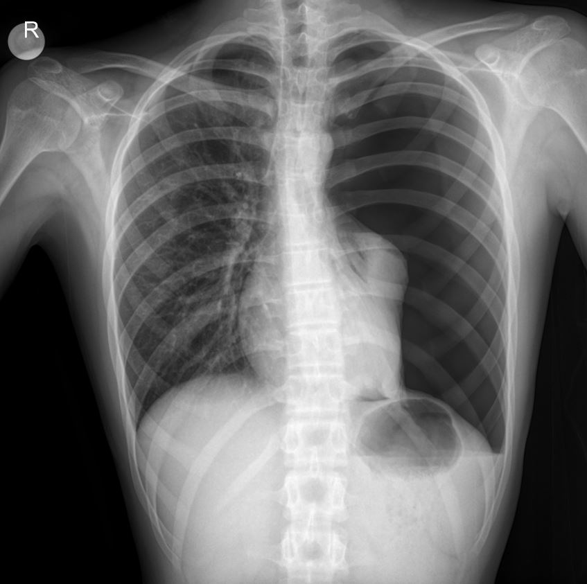
The provided chest X-ray image is highly demonstrative of a left total pneumothorax.
- Absence of Lung Markings: Crucially, there is a complete absence of vascular markings in the left hemithorax, indicating that the lung has fully collapsed.
- Visceral Pleural Line: A distinct, thin white line (indicated by the white line in the image) can be seen outlining the collapsed left lung, which is retracted towards the hilum. This line represents the visceral pleura.
- Hyperlucent Left Hemithorax: The left chest cavity appears hyperlucent (darker) compared to the right, due to the presence of air in the pleural space.
- Mediastinal Shift: While subtle, there may be a slight shift of the mediastinum (heart and trachea) towards the right, although this is less pronounced in a simple pneumothorax compared to a tension pneumothorax.
- Diaphragmatic Depression: The left hemidiaphragm may appear slightly depressed.
These radiological findings are pathognomonic for a total pneumothorax and correlate perfectly with the clinical finding of absent breath sounds on the left side.
Initial Management and Transfer
The patient was kept nil per os (NPO) and preoperative tests were initiated. He was immediately referred to the Pediatric Surgery department and subsequently transferred via ambulance to a specialized pulmonology center for definitive management. The patient has no history of smoking.
Please consult your national drug dosage guidelines or a licensed physician within the relevant department for specific medication types and dosages.
Conclusion
This case effectively illustrates the typical presentation and definitive radiological diagnosis of a left total pneumothorax in an adolescent. The rapid identification through physical examination and prompt confirmation with a chest X-ray were critical steps in initiating appropriate management. Early recognition and referral to specialized surgical care are paramount for optimal patient outcomes in such scenarios.
Disclaimer: This content is for educational and informational purposes only and should not be considered medical advice or a substitute for professional medical judgment, diagnosis, or treatment.

