The secondary follicle represents a crucial intermediate stage in the maturation of an ovarian follicle, bridging the gap between early development and the formation of a mature, ovulatory structure. This detailed diagram provides a microscopic view, highlighting the specific cellular layers and components that define this stage. Understanding the anatomy of a secondary follicle is fundamental to comprehending the complex process of folliculogenesis and its role in female reproduction.
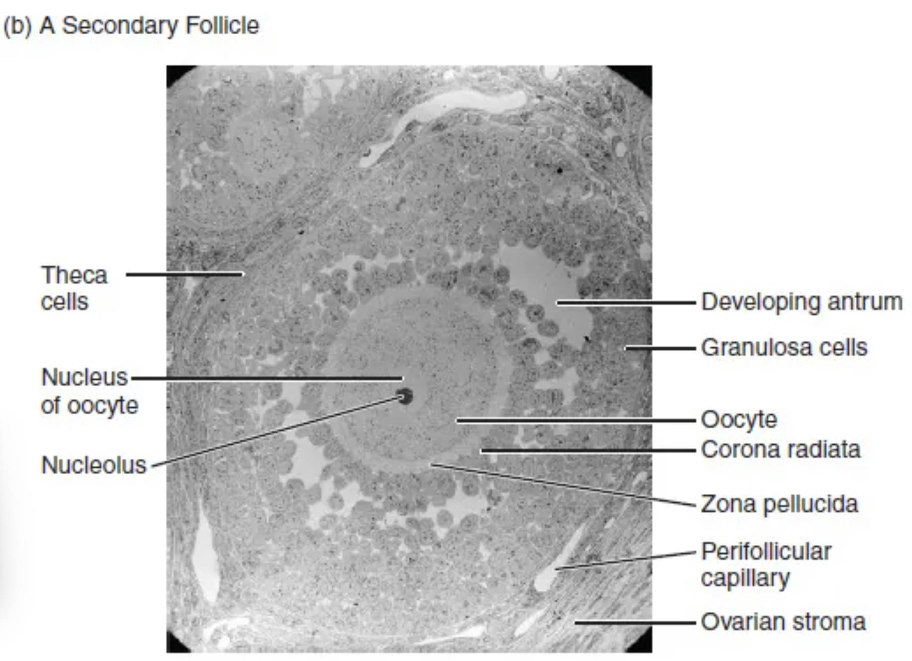
Theca cells: Theca cells are stromal cells that differentiate and form an outer layer surrounding the granulosa cells of the developing follicle. They are crucial for steroid hormone production, particularly synthesizing androgens that granulosa cells then convert into estrogens.
Nucleus of oocyte: The nucleus of the oocyte contains the genetic material (chromosomes) of the female gamete. At this stage, the oocyte is typically a primary oocyte arrested in prophase I of meiosis.
Nucleolus: The nucleolus is a dense structure found within the nucleus of the oocyte, primarily involved in ribosome synthesis and RNA production. Its prominent appearance indicates high metabolic activity within the growing oocyte.
Developing antrum: The developing antrum refers to the small, fluid-filled spaces that begin to coalesce within the granulosa cell layers of a secondary follicle. These spaces will eventually merge to form the single, large antrum characteristic of a tertiary follicle.
Granulosa cells: Granulosa cells are somatic cells that proliferate and form multiple layers around the oocyte in a secondary follicle. They are essential for providing nutrients to the oocyte and converting androgens from theca cells into estrogens.
Oocyte: The oocyte is the immature egg cell, housed within the center of the follicle. In a secondary follicle, the oocyte has grown significantly compared to earlier stages and is preparing for further meiotic progression.
Corona radiata: The corona radiata is the innermost layer of granulosa cells that directly surrounds the zona pellucida of the oocyte. These cells remain attached to the oocyte even after ovulation and play a role in fertilization.
Zona pellucida: The zona pellucida is a thick, glycoprotein layer that encapsulates the oocyte, acting as a protective barrier. It is vital for species-specific sperm binding and plays a role in preventing polyspermy.
Perifollicular capillary: Perifollicular capillaries are small blood vessels located in the stroma surrounding the follicle. They supply oxygen and nutrients to the developing follicle and transport hormones away from it.
Ovarian stroma: The ovarian stroma is the connective tissue framework of the ovary, providing structural support to the follicles and housing various cells, including undifferentiated mesenchymal cells that can differentiate into theca cells.
The Significance of the Secondary Follicle
The secondary follicle marks a pivotal stage in folliculogenesis, the process of ovarian follicle development, representing a significant progression from the simpler primary follicle. At this point, the follicle undergoes substantial growth and differentiation, laying the groundwork for further maturation into a dominant, ovulatory follicle. This stage is characterized by specific cellular proliferation and the emergence of new layers and structures, all working in concert to support the development of the oocyte.
The diagram provides a detailed histological view, allowing for a clear identification of the distinct cellular populations and their arrangements. The increase in granulosa cell layers and the appearance of theca cells are defining features, signaling an intensified level of metabolic activity and hormonal interplay. Understanding these anatomical changes is crucial for comprehending the dynamics of the menstrual cycle and the hormonal environment required for successful reproduction.
Key features of a secondary follicle include:
- Multiple layers of proliferating granulosa cells.
- The differentiation of surrounding theca cells.
- The continued growth of the oocyte and thickening of the zona pellucida.
- The initial formation of fluid-filled spaces, indicative of a developing antrum.
Disruptions at this stage can impact the quality of the oocyte and potentially lead to fertility challenges, highlighting the importance of normal secondary follicle development.
Cellular Interactions and Hormonal Synthesis
Within a secondary follicle, the communication between different cell types is remarkably sophisticated. The granulosa cells, now forming multiple layers around the growing oocyte, play a critical role in providing essential nutrients and growth factors to the oocyte. Crucially, they also initiate the conversion of androgens into estrogens, a process driven by the enzyme aromatase. These androgens are primarily supplied by the surrounding theca cells, which differentiate from the ovarian stroma. Theca cells are highly vascularized, allowing them to effectively take up cholesterol precursors and synthesize androgens under the influence of Luteinizing Hormone (LH). This two-cell, two-gonadotropin theory of estrogen synthesis is fundamental to ovarian physiology.
The oocyte itself, with its prominent nucleus and nucleolus, continues to grow and prepare for the eventual completion of meiosis. The zona pellucida, a robust extracellular matrix, becomes thicker, serving as a protective coat and playing a vital role in species-specific sperm recognition during fertilization. Small, fluid-filled spaces begin to appear between the granulosa cells, marking the very early stages of antrum formation that will become a defining feature of the tertiary follicle. The surrounding perifollicular capillaries ensure adequate blood supply, delivering hormones and nutrients while removing waste products. This intricate cellular organization and coordinated hormonal activity within the secondary follicle are paramount for the progression towards ovulation and the potential for successful reproduction.
In conclusion, the secondary follicle is a dynamic and functionally significant stage in ovarian development, characterized by cellular proliferation, hormonal synthesis, and intricate cell-to-cell communication. Its detailed structure provides a window into the complex biological processes that prepare an oocyte for maturation and potential fertilization. A comprehensive understanding of the secondary follicle’s anatomy and physiology is indispensable for grasping female reproductive health, fertility treatments, and the broader field of endocrinology.

