The male reproductive system’s efficiency hinges on the precise functioning of its core components, particularly the testes, which are housed within the scrotum. This diagram provides a comprehensive anterior view, dissecting the layers of the scrotum to reveal the intricate structures within, including the testes, epididymides, and the vital elements of the spermatic cord. Understanding this complex arrangement is fundamental to grasping the mechanisms of spermatogenesis, temperature regulation, and overall male reproductive health.
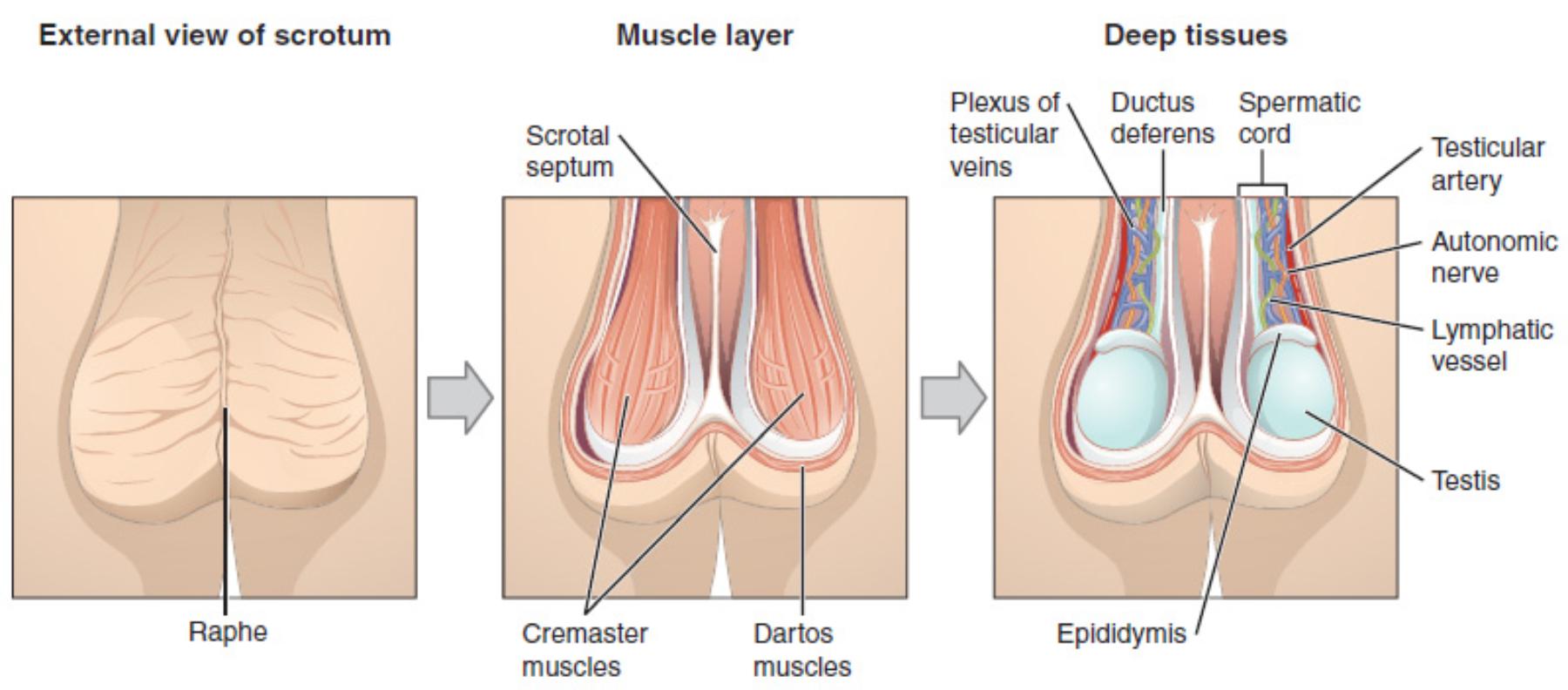
Deconstructing the Scrotal and Testicular Structures
External view of scrotum: This initial image shows the superficial appearance of the scrotum, which is a sac of skin and superficial fascia. It is located inferior to the pubic symphysis and anterior to the anus, serving as the protective external housing for the testes.
Raphe: A distinct ridge or seam visible on the external surface of the scrotum, running longitudinally down the midline. This anatomical landmark is a remnant of the fusion of the two scrotal sacs during embryonic development.
Muscle layer: This cross-sectional view reveals the muscular layers beneath the skin of the scrotum. These muscles play a crucial role in thermoregulation, maintaining the optimal temperature required for spermatogenesis.
Scrotal septum: An internal wall or partition of fascia and muscle that divides the scrotum into two compartments. Each compartment houses one testis, epididymis, and the associated spermatic cord, preventing the spread of infection between the two sides.
Cremaster muscles: Striated skeletal muscles located within the spermatic cord and surrounding the testes. They are responsible for elevating the testes closer to the body in response to cold temperatures or sexual arousal, a reflex known as the cremasteric reflex.
Dartos muscles: A layer of smooth muscle located in the superficial fascia of the scrotum, directly beneath the skin. Contraction of the dartos muscle causes the scrotal skin to wrinkle and thicken, reducing surface area and heat loss, while relaxation has the opposite effect.
Deep tissues: This deepest cross-section illustrates the internal structures, including the testes, epididymides, and the complex components of the spermatic cord. These tissues are vital for sperm production, maturation, and transport.
Plexus of testicular veins: Also known as the pampiniform plexus, this is a network of many small veins that drain the testis and epididymis. It plays a critical role in cooling arterial blood before it reaches the testes, acting as a countercurrent heat exchanger.
Ductus deferens: A muscular tube that transports mature sperm from the epididymis to the ejaculatory duct. It is a key component of the spermatic cord and contracts forcefully during ejaculation to propel sperm.
Spermatic cord: A cord-like structure in the male that extends from the abdomen into the scrotum. It contains the ductus deferens, testicular artery, pampiniform plexus of veins, nerves, and lymphatic vessels, all bundled together.
Testicular artery: An artery that supplies oxygenated blood to the testis and epididymis. It branches off the abdominal aorta and descends into the scrotum as a component of the spermatic cord.
Autonomic nerve: Nerves that innervate the smooth muscles of the blood vessels and ducts within the testis and epididymis, regulating processes such as blood flow and ejaculation. They are part of the autonomic nervous system.
Lymphatic vessel: Vessels that drain lymph from the testes and epididymides. These vessels are important for immune surveillance and fluid balance, eventually returning lymph to the systemic circulation.
Testis: One of two oval-shaped male gonads located in the scrotum. The testis is the primary male reproductive organ, responsible for spermatogenesis (sperm production) and the synthesis of male sex hormones, primarily testosterone.
Epididymis: A comma-shaped organ located posterior to each testis. It serves as a site for sperm maturation and storage, where sperm gain motility and the ability to fertilize an ovum.
The Scrotum and Testes: Guardians of Spermatogenesis
The scrotum and its contents, primarily the testes, represent a cornerstone of the male reproductive system. This intricate anatomical arrangement, clearly depicted in the diagram, is meticulously designed to optimize spermatogenesis—the process of sperm production—which requires a specific temperature range slightly cooler than the core body temperature. The scrotum acts as a dynamic environmental regulator, adapting to external conditions to maintain this critical thermal environment.
The outermost layer, the skin of the scrotum, is characterized by the raphe, a visible midline seam. Beneath this lies a crucial muscular layer comprising the dartos and cremaster muscles. The dartos muscle, a smooth muscle, contracts to wrinkle and thicken the scrotal skin in cold conditions, reducing heat loss. Conversely, the cremaster muscles, skeletal muscles within the spermatic cord, elevate the testes closer to the body when it’s cold or during sexual arousal, further contributing to temperature regulation. This dual muscular control allows for precise thermoregulation, creating an optimal microclimate for sperm development within the testes.
Within the protective confines of the scrotum, separated by the scrotal septum, reside the testes and epididymides. The testes are the primary male gonads, responsible for both sperm production and the synthesis of testosterone. Attached to each testis is the epididymis, a coiled tube where sperm mature and are stored until ejaculation. Accompanying these structures are the components of the spermatic cord, which includes the ductus deferens (transporting sperm), the testicular artery (supplying blood), the pampiniform plexus of testicular veins (a heat exchanger), autonomic nerves, and lymphatic vessels. This complex vascular and neural network underscores the sophisticated physiological demands of male reproduction.
Decoding the Anatomy of the Scrotum and Testes
The male reproductive system’s ability to produce viable sperm relies heavily on the specific anatomical and physiological conditions maintained within the scrotum. This anterior view diagram provides an excellent step-by-step breakdown of the layers and intricate structures that constitute the scrotum and its vital contents, primarily the testes. Understanding this organizational complexity is paramount for grasping male fertility, hormone production, and the numerous clinical conditions that can affect this crucial region.
Beginning with the external appearance, the scrotum is a cutaneous fibromuscular sac that encases the testes. Its external midline is marked by the raphe, a visible ridge that signifies embryonic fusion. Directly beneath the skin lies the dartos muscle, a layer of smooth muscle within the superficial fascia. Contraction of the dartos muscle causes the scrotal skin to wrinkle and contract, reducing the surface area and pulling the testes closer to the body to conserve heat. Conversely, relaxation allows the scrotum to hang lower, facilitating heat dissipation. This thermoregulatory function is critical because spermatogenesis, the process of sperm production, is highly temperature-sensitive and requires an environment approximately 2-3°C cooler than the core body temperature.
Further dissecting the scrotal layers reveals the cremaster muscles, which are extensions of the internal oblique abdominal muscle. These striated skeletal muscles are housed within the spermatic cord and can contract reflexively (the cremasteric reflex) to quickly elevate the testes towards the inguinal canal in response to cold or sexual arousal. Internally, the scrotum is divided into two compartments by the scrotal septum, preventing unilateral infections from easily spreading to the other side. Within these compartments are the testes, the primary male gonads responsible for both spermatogenesis and androgen production, predominantly testosterone. Each testis is intimately associated with an epididymis, a comma-shaped structure where sperm undergo their final maturation process and are stored before ejaculation.
The diagram also highlights the vital components of the spermatic cord, which descends from the inguinal canal into the scrotum. This cord is a conduit for various structures essential for testicular function, including the ductus deferens (vas deferens), which transports mature sperm; the testicular artery, providing oxygenated blood; the pampiniform plexus of veins, a unique venous network that acts as a countercurrent heat exchanger to cool the arterial blood flowing to the testes; autonomic nerves for regulation; and lymphatic vessels for fluid drainage and immune surveillance. The intricate arrangement of these structures underscores the sophistication of the male reproductive system in ensuring optimal conditions for fertility.
Conclusion
This detailed anterior view of the scrotum and testes illuminates the complex anatomical and physiological adaptations that ensure the viability of male reproduction. From the external raphe and the thermoregulatory muscles (dartos and cremaster) to the internal testes, epididymides, and the vital components of the spermatic cord, each structure plays a critical role. A thorough understanding of this intricate design is essential for comprehending the mechanisms of spermatogenesis, hormonal regulation, and for diagnosing and managing conditions affecting male reproductive health.

