The male reproductive system is a complex network of organs and ducts meticulously designed for the production, maturation, and delivery of sperm, alongside the synthesis of male sex hormones. This detailed diagram offers a multifaceted view, illustrating both the external genitalia with variations in penile anatomy and a comprehensive lateral view of the internal structures. Understanding the intricate relationships between the testes, epididymides, various glands, and associated ducts is fundamental to comprehending male reproductive physiology and potential clinical implications.
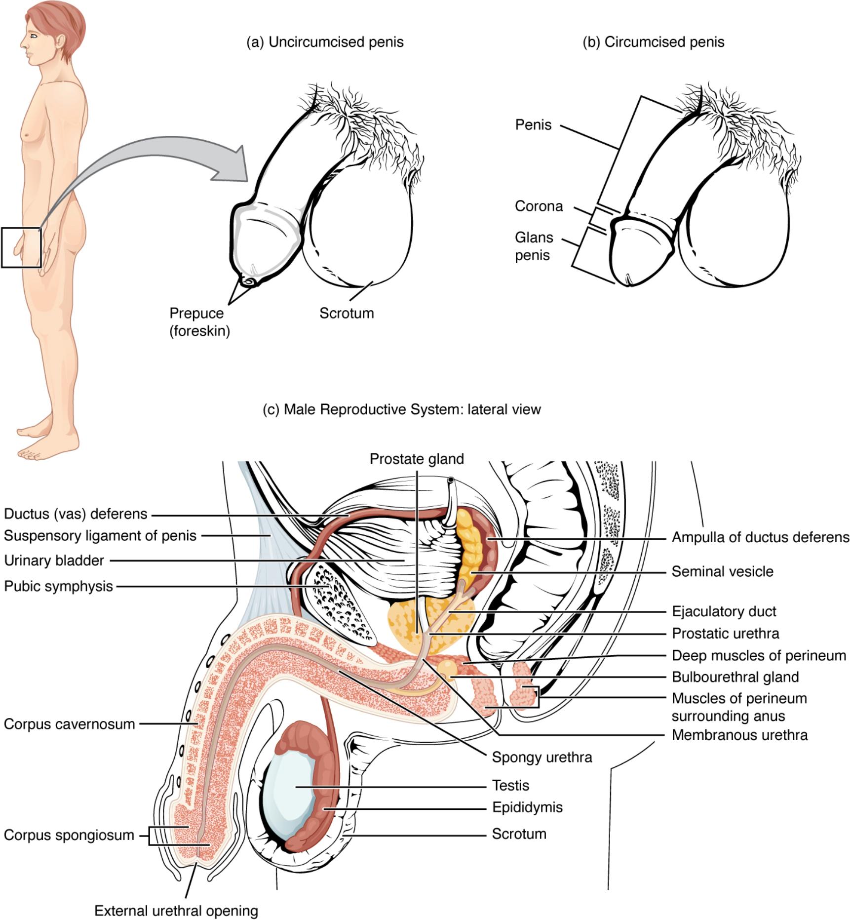
Exploring the Structures of the Male Reproductive System
a) Uncircumcised penis: This illustration depicts the external appearance of an uncircumcised penis, characterized by the presence of a prepuce, or foreskin, covering the glans penis. This natural anatomical state is found in males who have not undergone the surgical procedure of circumcision.
b) Circumcised penis: This illustration shows the external appearance of a circumcised penis, where the prepuce (foreskin) has been surgically removed, leaving the glans penis exposed. This is a common practice in many cultures and for various medical or religious reasons.
Penis: The primary external male copulatory organ, through which both urine and semen are expelled from the body. It consists of a shaft and the glans penis, and its internal structure involves erectile tissues like the corpora cavernosa and corpus spongiosum.
Corona: The crown-like ridge located at the base of the glans penis, separating the glans from the shaft. It is a highly sensitive area.
Glans penis: The enlarged, rounded tip of the penis, which is richly innervated and highly sensitive. In an uncircumcised penis, it is covered by the prepuce.
Prepuce (foreskin): A retractable fold of skin that covers and protects the glans penis in an uncircumcised male. It can be moved back to expose the glans.
Scrotum: A sac of skin and superficial fascia suspended inferior to the pubic symphysis and anterior to the anus. It contains the testes, epididymides, and the initial parts of the spermatic cords, maintaining a temperature suitable for spermatogenesis.
c) Male Reproductive System: lateral view: This comprehensive cross-sectional diagram provides a lateral internal view of the male reproductive organs, showcasing their anatomical relationships within the pelvic cavity and externally. It highlights the intricate network of ducts and glands involved in semen production and transport.
Ductus (vas) deferens: A muscular tube that transports sperm from the epididymis to the ejaculatory duct. It is a key component of the spermatic cord and contracts forcefully during ejaculation to propel sperm.
Suspensory ligament of penis: A fibrous band that extends from the pubic symphysis to the dorsal surface of the penis. It helps to support the weight of the penis and anchors it to the pelvis.
Pubic symphysis: A cartilaginous joint located anteriorly between the two pubic bones of the pelvis. It provides a point of attachment for various pelvic structures, including the ligaments supporting the penis.
Urinary bladder: A muscular sac that stores urine received from the kidneys via the ureters. While part of the urinary system, its anatomical proximity and the passage of the urethra through the penis mean it is often visualized in diagrams of the male reproductive system.
Corpus cavernosum: One of two large, dorsal columns of erectile tissue within the shaft of the penis. These spongy tissues fill with blood during sexual arousal, causing the penis to become erect.
Corpus spongiosum: A single, ventral column of erectile tissue within the penis, which surrounds the urethra. It helps to keep the urethra open during erection, preventing its compression.
External urethral opening: The distal opening of the urethra located at the tip of the glans penis. It serves as the exit point for both urine and semen.
Prostate gland: A walnut-sized gland located inferior to the urinary bladder and surrounding the prostatic urethra. It secretes a milky, slightly acidic fluid containing citrate (nutrient for sperm), enzymes, and prostate-specific antigen (PSA), contributing significantly to semen volume and viability.
Seminal vesicle: One of two sac-like glands located posterior to the urinary bladder. These glands secrete a yellowish, alkaline fluid rich in fructose (energy for sperm), prostaglandins, and clotting factors, accounting for about 60% of semen volume.
Ampulla of ductus deferens: The enlarged terminal portion of the ductus deferens, located just before it joins the seminal vesicle duct to form the ejaculatory duct. It serves as a temporary storage site for sperm prior to ejaculation.
Ejaculatory duct: Formed by the union of the ductus deferens ampulla and the seminal vesicle duct. It passes through the prostate gland and empties into the prostatic urethra, allowing sperm and seminal fluid to mix.
Prostatic urethra: The portion of the urethra that passes through the prostate gland. It receives ejaculatory ducts and ducts from the prostate gland.
Deep muscles of perineum: A group of muscles located in the pelvic floor, essential for supporting pelvic organs and playing a role in micturition and ejaculation.
Bulbourethral gland: Also known as Cowper’s gland, one of two small glands located inferior to the prostate gland, at the base of the penis. They secrete a clear, alkaline mucus into the spongy urethra before ejaculation, which lubricates the urethra and neutralizes any residual acidic urine.
Muscles of perineum surrounding anus: Refers to the muscles of the pelvic floor, including the external anal sphincter, which are crucial for continence and also support the pelvic organs.
Membranous urethra: The short, narrow portion of the urethra that passes through the deep muscles of the perineum. It connects the prostatic urethra to the spongy urethra.
Spongy urethra: The longest part of the urethra, extending through the corpus spongiosum of the penis to the external urethral opening. It transports urine and semen out of the body.
Testis: One of two oval-shaped male gonads located in the scrotum. The testes are responsible for spermatogenesis (sperm production) and the synthesis of male sex hormones, primarily testosterone.
Epididymis: A comma-shaped organ located posterior to each testis. It serves as a site for sperm maturation and storage, where sperm gain motility and the ability to fertilize an ovum.
The male reproductive system is a marvel of biological engineering, specifically designed to perpetuate the species through the production and delivery of male gametes (sperm) and the synthesis of essential sex hormones. This intricate system comprises both external genitalia and a complex network of internal ducts and glands, all working in concert. The provided diagram offers a comprehensive anatomical overview, highlighting the key structures involved in these vital processes.
The external structures, as depicted in figures (a) and (b), primarily include the penis and the scrotum. The penis, the male copulatory organ, facilitates the transfer of semen into the female reproductive tract. Its anatomical variations, specifically the presence or absence of the prepuce (foreskin), are illustrated, distinguishing between uncircumcised and circumcised penises. The glans penis, corona, and external urethral opening are also clearly labeled, showcasing the distal components of this organ. The scrotum, a protective sac, houses the testes at a temperature slightly lower than body temperature, an optimal condition for spermatogenesis.
Figure (c) provides a detailed lateral view of the internal male reproductive organs, revealing the complex plumbing and glandular contributions essential for fertility. Key internal structures include:
- Testes: The primary male gonads, responsible for sperm production and hormone synthesis.
- Epididymis: A coiled tube where sperm mature and are stored.
- Ductus (vas) deferens: A muscular tube that transports mature sperm from the epididymis towards the ejaculatory duct.
- Seminal Vesicles and Prostate Gland: Accessory glands that contribute fluids to sperm, forming semen. These secretions provide nutrients, buffers, and substances that aid in sperm motility and viability.
- Urethra: The terminal duct that carries both urine and semen out of the body.
The journey of sperm from its production in the testes to its expulsion involves a precise sequence of events and contributions from various glands. Sperm mature in the epididymis and are then transported through the ductus deferens. Along this pathway, they mix with fluids from the seminal vesicles, prostate gland, and bulbourethral glands, collectively forming semen. This complex fluid mixture then travels through the ejaculatory ducts and the urethra, ready for ejaculation.
Anatomy and Function of the Male Reproductive System
The male reproductive system is a remarkable biological apparatus essential for reproduction and the production of male hormones. This system is composed of primary and accessory organs, each with a specialized role in ensuring the viability and delivery of sperm. A comprehensive understanding of its anatomy and physiology is crucial for appreciating male health and fertility.
The primary reproductive organs are the testes, located within the scrotum, an external sac that provides a cooler environment vital for spermatogenesis (sperm production). Each testis is responsible for generating millions of sperm daily and for synthesizing androgens, primarily testosterone, which drives male secondary sexual characteristics and reproductive function. From the testes, immature sperm are transported to the epididymis, a highly coiled tube located posterior to each testis. Here, sperm undergo a crucial maturation process, gaining motility and the capacity to fertilize an ovum.
Following maturation, sperm are stored in the epididymis until ejaculation. During sexual arousal, sperm are propelled from the epididymis through the ductus deferens (also known as the vas deferens). Each ductus deferens extends superiorly into the pelvic cavity, loops over the urinary bladder, and widens to form the ampulla. The ampulla then joins with the duct of the seminal vesicle to form the ejaculatory duct. These ducts converge within the prostate gland, ultimately emptying into the prostatic urethra. Along this pathway, accessory glands contribute essential fluids to create semen.
The accessory glands include the seminal vesicles, prostate gland, and bulbourethral glands. The seminal vesicles secrete an alkaline fluid rich in fructose, a sugar that provides energy for sperm motility, along with prostaglandins and clotting factors. The prostate gland, situated inferior to the bladder, produces a milky, slightly acidic fluid containing citrate (another sperm nutrient), enzymes, and prostate-specific antigen (PSA), which helps to liquefy the semen after ejaculation. Finally, the bulbourethral glands secrete a clear, alkaline mucus prior to ejaculation, lubricating the urethra and neutralizing any residual acidic urine, thereby creating a more favorable environment for sperm passage. The combined secretions from these glands mix with sperm to form semen, a complex fluid designed to protect, nourish, and transport sperm effectively through the female reproductive tract. The semen is ultimately expelled from the body through the urethra, which traverses the length of the penis. The penis itself, composed of erectile tissues like the corpora cavernosa and corpus spongiosum, facilitates copulation through its ability to become erect upon sexual stimulation.
Conclusion
The detailed anatomical representation of the male reproductive system, encompassing both external and internal structures, provides invaluable insight into its intricate design and critical functions. From the testes’ role in sperm and hormone production to the sophisticated network of ducts and glands that facilitate sperm maturation and semen formation, each component plays an indispensable part in male fertility and overall physiological well-being. A thorough understanding of these structures and their coordinated functions is essential for grasping the complexities of human reproduction and addressing various health concerns related to the male reproductive system.

