Male Karyotype: A Comprehensive Guide to Chromosomal Structure and Genetic Organization
This image presents a karyotype, a visual representation of a male individual’s complete set of chromosomes, organized by size and banding patterns. This essential diagnostic tool allows for the analysis of an individual’s chromosomal complement, revealing the intricate organization of their genetic material. Understanding karyotypes is fundamental to comprehending genetic health and the basis of inherited traits.
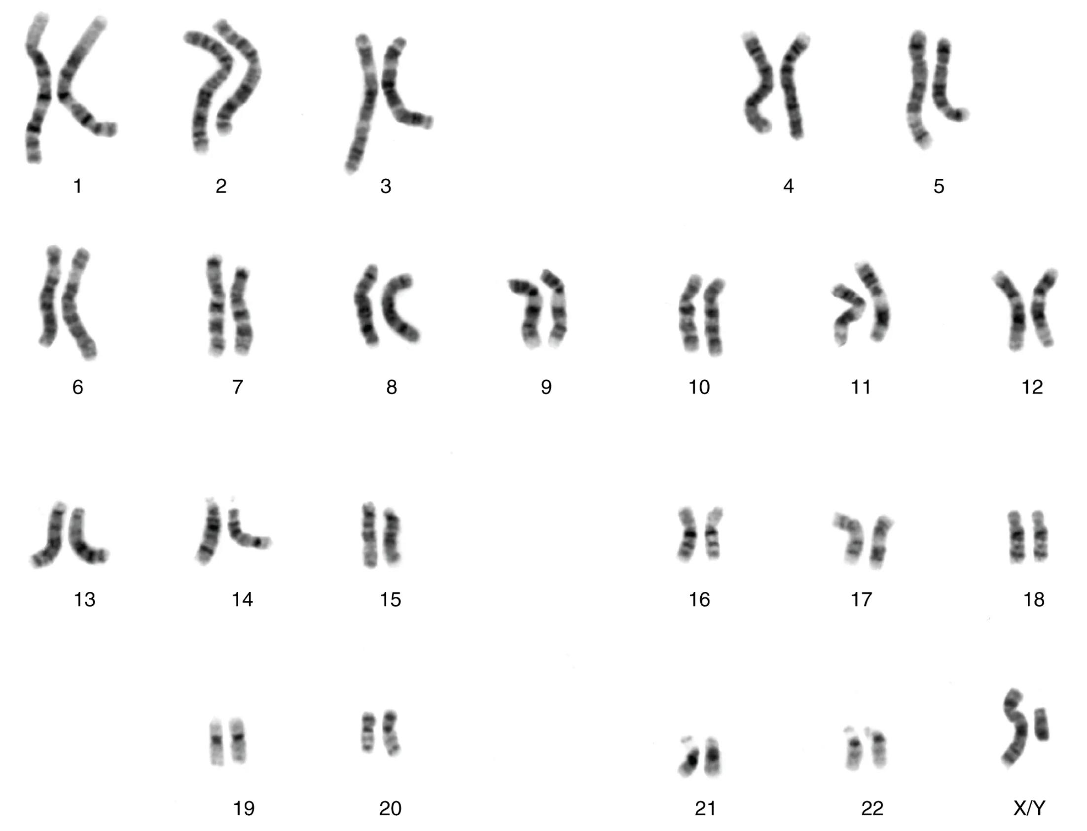
Chromosomes 1-22: These are the autosomes, non-sex chromosomes that carry the majority of an individual’s genetic information. Each pair, from 1 to 22, demonstrates a remarkably consistent banding pattern between the two homologous chromosomes, signifying the identical organization of thousands of genes. These chromosomes are responsible for coding for most of the body’s traits and functions, ranging from physical characteristics to metabolic processes.
X/Y: This pair represents the sex chromosomes, which determine an individual’s biological sex. In this male karyotype, an X chromosome and a Y chromosome are present. The X chromosome is significantly larger and contains many genes essential for both male and female development, while the Y chromosome is smaller and primarily carries genes crucial for male sex determination and development. The distinct size and gene content of the X and Y chromosomes are the only exceptions to the otherwise nearly identical pairing seen in autosomes.
A karyotype is an organized profile of a person’s chromosomes, arranged in homologous pairs from largest to smallest. This specific image showcases the chromosomal complement of a male, characterized by the presence of both an X and a Y sex chromosome. The distinctive banding patterns visible on each chromosome are crucial, as they represent regions of DNA that stain differently, providing a unique genetic “barcode” for each chromosome. These bands are not merely decorative; they reflect the underlying sequence and organization of genes, with each band containing hundreds to thousands of genes.
The principle of homologous pairing is vividly demonstrated here. For autosomes (pairs 1-22), the two chromosomes within each pair exhibit nearly identical banding patterns. This visual congruency confirms that these pairs carry the same set of genes, arranged in the same order, albeit with potentially different alleles (variations of a gene) inherited from each parent. The integrity of these paired structures is vital for normal cellular function and inheritance. Any significant deviation in number or structure can have profound implications for an individual’s health and development.
- The meticulous arrangement of chromosomes in a karyotype allows for the detection of chromosomal abnormalities, such as aneuploidies (abnormal number of chromosomes) or structural rearrangements (deletions, duplications, translocations). These anomalies are often associated with genetic disorders and developmental challenges.
This detailed examination of a male karyotype provides a foundational understanding of human genetics. It underscores how the precise organization and integrity of our chromosomes are paramount for health, development, and the inheritance of traits across generations. The unique characteristics of the sex chromosomes further highlight the genetic basis of sexual differentiation.
The study of the human karyotype is a cornerstone of genetics and medical diagnostics. Each of the 23 pairs of chromosomes, comprising 22 pairs of autosomes and one pair of sex chromosomes, holds a vast amount of genetic information, dictating everything from our physical attributes to our susceptibility to certain diseases. In a normal human male, the presence of an XY sex chromosome pair, as depicted, is the defining characteristic differentiating it from a female karyotype (XX). The remarkable consistency in banding patterns within autosomal pairs signifies an orderly and redundant genetic blueprint, where each chromosome in a pair carries homologous genes. This precise organization ensures that essential proteins and cellular functions are adequately coded for.
Variations in these banding patterns or chromosome numbers can indicate significant genetic conditions. For instance, trisomy 21, commonly known as Down syndrome, is characterized by an extra copy of chromosome 21. Other conditions, such as Klinefelter syndrome (XXY in males) or Turner syndrome (XO in females), arise from abnormalities in the sex chromosomes. The ability to visualize and analyze these chromosomal structures through karyotyping is invaluable for prenatal diagnosis, identifying causes of developmental delays, and guiding reproductive counseling. It provides a macroscopic view of the genome, complementing more granular genetic analyses like DNA sequencing.
Understanding the chromosomal complement is not merely an academic exercise; it has profound clinical applications. Genetic counselors utilize karyotype analysis to assess risks for inherited disorders, while oncologists may use it to identify chromosomal rearrangements in cancer cells, which can guide treatment strategies. The image serves as a powerful reminder of the intricate biological architecture that underpins human life, where each chromosome plays a critical role in the symphony of genetic expression. The delicate balance and precise organization within this microscopic world are fundamental to health and development.

