This comprehensive diagram illustrates the intricate sequence of events encompassing ovulation, fertilization, pre-embryonic development, and implantation within the female reproductive system. These critical stages, occurring within approximately one week, transform an unfertilized oocyte into a blastocyst poised for uterine attachment. Understanding this timeline and the anatomical locations of each event is fundamental to comprehending early human development and potential reproductive challenges.
Unfertilized Oocyte: An Unfertilized Oocyte is the mature female gamete, released during ovulation and awaiting fertilization. It is typically surrounded by the zona pellucida and cumulus cells, and it is arrested at metaphase II of meiosis.
Fertilized Oocyte: A Fertilized Oocyte, also known as a zygote, is the diploid cell formed by the fusion of a sperm and an egg. This event marks the genetic beginning of a new individual and initiates the process of embryonic development.
Two-cell stage: The Two-cell stage is the result of the first cleavage division of the zygote, typically occurring around 24-30 hours after fertilization. Each of these daughter cells is called a blastomere.
Four-cell stage: The Four-cell stage is achieved after the second cleavage division, producing four blastomeres. This developmental milestone is usually reached approximately 40-50 hours post-fertilization.
Eight-cell stage: The Eight-cell stage is the outcome of the third cleavage division, resulting in eight blastomeres. This stage is generally observed around 60-72 hours after fertilization.
Morula (16 cells): The Morula is a solid ball of 12-32 blastomeres, resembling a mulberry, that forms by day 3-4 of development. It is still enclosed within the zona pellucida.
Trophoblast: The Trophoblast is the outer layer of cells of the blastocyst, essential for mediating implantation into the uterine wall. These cells will later contribute to the formation of the placenta.
Inner cell mass: The Inner cell mass is a cluster of pluripotent cells located within the blastocyst, destined to give rise to the embryo proper. It is the source of all the tissues and organs of the developing fetus.
Blastocoel: The Blastocoel is a fluid-filled cavity within the blastocyst, formed through the active transport of sodium ions into the morula, drawing water in by osmosis. This cavity is crucial for the expansion and subsequent hatching of the blastocyst.
Blastocyst (70–100 cells): A Blastocyst is a highly organized embryonic stage, characterized by the presence of the trophoblast, inner cell mass, and blastocoel. It typically forms around day 5-6 post-fertilization and is the stage at which implantation occurs.
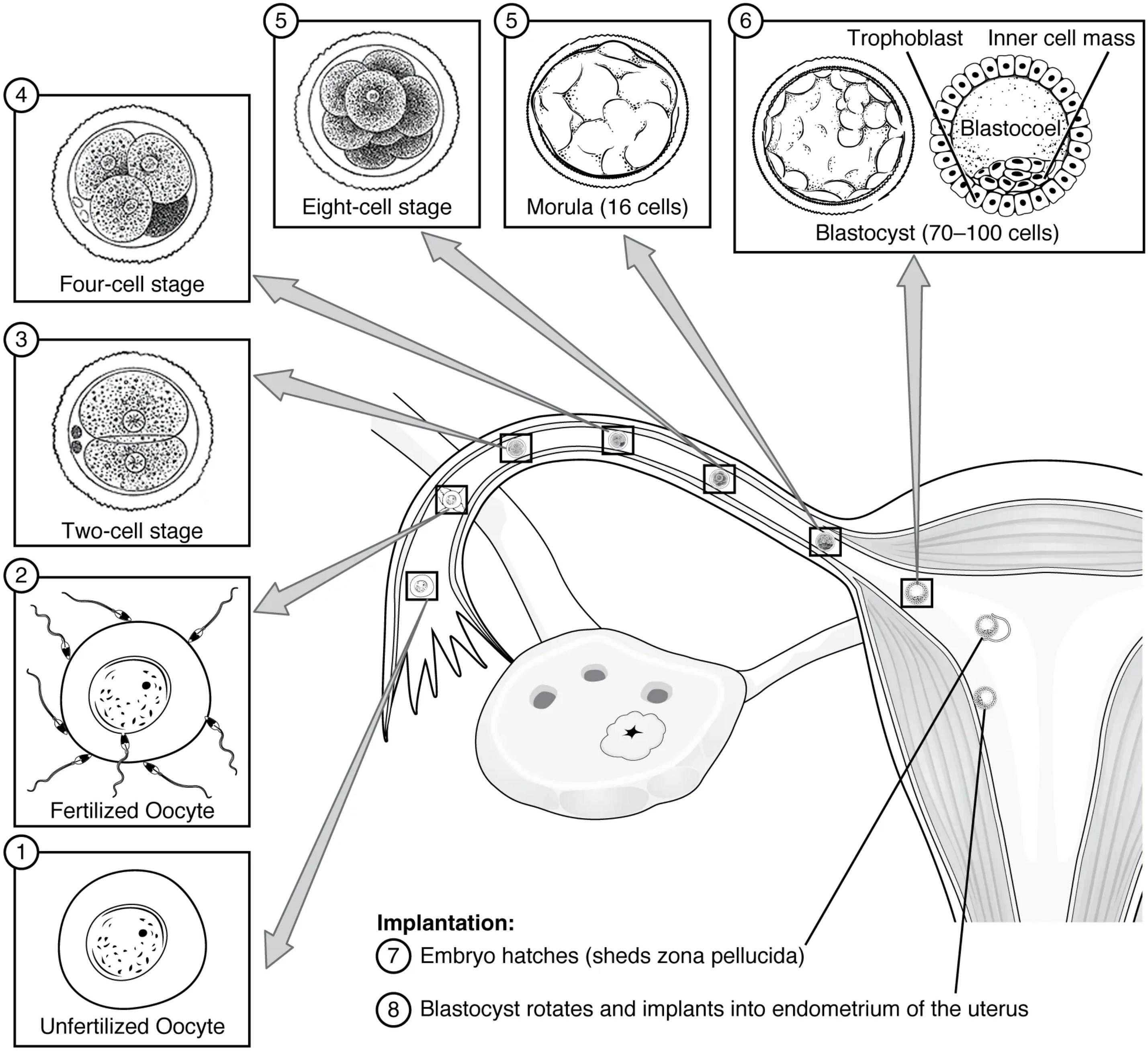
The remarkable journey of early human development commences with an Unfertilized Oocyte (1), typically released from the ovary during ovulation. This oocyte then travels into the fallopian tube, where fertilization occurs, transforming it into a Fertilized Oocyte (2) or zygote. This pivotal event, usually taking place in the ampulla of the fallopian tube, marks the initiation of a series of rapid mitotic divisions known as pre-embryonic development or cleavages.
As the fertilized oocyte begins its descent towards the uterus, it undergoes sequential divisions, progressing from the Two-cell stage (3), to the Four-cell stage (4), and then to the Eight-cell stage (5). These divisions, while increasing the number of cells (blastomeres), do not increase the overall size of the conceptus, as they occur within the confines of the zona pellucida. This conservation of volume is essential for the embryo to traverse the narrow lumen of the fallopian tube.
Further divisions and compaction lead to the formation of the Morula (16 cells) (5) by about day 3-4 post-fertilization. As the morula enters the uterine cavity, it transforms into a Blastocyst (70–100 cells) (6) by day 5-6. This blastocyst is a complex structure featuring an Inner cell mass (which will form the embryo), an outer Trophoblast layer (for implantation and placental development), and a fluid-filled Blastocoel.
The final, crucial step is Implantation, where the embryo hatches (sheds zona pellucida) (7), freeing itself to directly interact with the uterine lining. The blastocyst rotates and implants into the endometrium of the uterus (8) approximately 6-7 days post-fertilization. This precise timing and location are critical for establishing a successful pregnancy, highlighting the intricate orchestration of biological events in early human development.
The Stages of Early Human Development
The first week of human development, often referred to as pre-embryonic development, is a highly dynamic period that lays the foundation for all subsequent growth. This phase is characterized by rapid cellular division, migration, and differentiation, occurring within specific anatomical locations of the female reproductive system. A clear understanding of these events is crucial for recognizing normal development and potential deviations.
From Fertilization to Blastocyst Formation
Following ovulation, the Unfertilized Oocyte is typically swept into the fallopian tube, where it may encounter sperm. Fertilization usually occurs in the ampulla of the fallopian tube, resulting in the Fertilized Oocyte (zygote). This single cell immediately begins a series of mitotic divisions, known as cleavages. The Two-cell stage is followed by the Four-cell stage, and then the Eight-cell stage, all within the confines of the zona pellucida, which acts as a protective barrier and prevents premature implantation. This sequential division culminates in the formation of the Morula by day 3-4.
Blastocyst Maturation and Implantation
As the Morula continues its journey towards the uterus, it transforms into a Blastocyst by day 5-6. This transition involves the formation of the Blastocoel, a fluid-filled cavity, and the differentiation of cells into the Inner cell mass and the surrounding Trophoblast. The Trophoblast cells are instrumental for the upcoming Implantation process. Before implantation can occur, the Embryo hatches (sheds zona pellucida), allowing direct contact between the Trophoblast and the uterine lining. Subsequently, the blastocyst rotates and implants into the endometrium of the uterus, typically on day 6 or 7. This critical event establishes the connection between the developing embryo and the maternal circulation, marking the formal beginning of pregnancy.

