This comprehensive diagram illustrates the key stages of In Vitro Fertilization (IVF), a sophisticated assisted reproductive technology that offers hope to individuals and couples facing infertility. The process involves the careful collection of eggs from the ovaries, their fertilization with sperm in a controlled laboratory setting (a petri dish), and the subsequent transfer of viable embryos into the uterus. This detailed visual guide demystifies each step, from gamete retrieval to successful implantation.
Egg (oocyte): An egg (oocyte) is the female reproductive cell, typically released from the ovary during ovulation. In IVF, multiple oocytes are stimulated to mature and are then carefully collected for fertilization outside the body.
Catheter: A catheter is a thin, flexible tube used in medical procedures for various purposes, including the aspiration of eggs from ovarian follicles during oocyte retrieval. Its precise manipulation is crucial for successful collection while minimizing trauma.
Fertilized zygote: A fertilized zygote is the single cell formed when a sperm successfully fuses with an egg, containing the complete diploid set of chromosomes. In IVF, these zygotes are closely monitored for initial development before embryo transfer.
Implanted zygote: An implanted zygote refers to the early embryo (at the blastocyst stage) that has successfully attached to and embedded itself within the lining of the uterus. Successful implantation is a critical step for the continuation of pregnancy.
Endometrium: The endometrium is the inner lining of the uterus, which thickens each month in preparation for potential pregnancy. It is the site where the implanted zygote (embryo) will embed and develop.
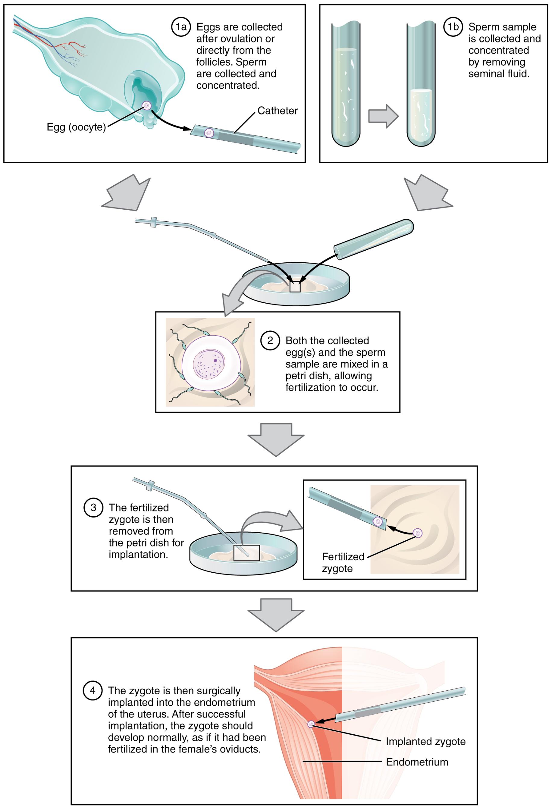
The journey of IVF begins with meticulous preparation, as shown in step 1a, where eggs (oocytes) are carefully collected after ovulation or directly from the ovarian follicles using a catheter. Concurrently, in step 1b, a sperm sample is collected and processed to concentrate viable sperm by removing seminal fluid. These initial steps are fundamental to ensuring the availability of high-quality gametes for the fertilization process.
Following gamete collection, step 2 depicts the crucial phase where both the collected egg(s) and the prepared sperm sample are combined in a petri dish. This controlled environment allows for fertilization to occur spontaneously, mimicking the natural process but under expert supervision. The successful fusion of a sperm with an egg results in the formation of a fertilized zygote.
Once fertilization has taken place and the zygote begins its initial stages of development, it is carefully removed from the petri dish, as illustrated in step 3. This delicate manipulation is performed with specialized instruments to prepare the early embryo for its transfer back into the female reproductive tract. The quality and developmental stage of the fertilized zygote are critical factors considered before proceeding to implantation.
Finally, in step 4, the zygote (now typically an embryo at a more advanced stage of development) is surgically implanted into the endometrium of the uterus. This final step is meticulously performed to maximize the chances of successful implantation and, subsequently, a healthy pregnancy. If successful, the implanted zygote should develop normally, just as if fertilization had occurred naturally within the female’s oviducts, culminating in a successful gestation.
Understanding the IVF Process: A Detailed Insight
In vitro fertilization (IVF) has revolutionized the treatment of infertility, offering a pathway to parenthood for many who previously had limited options. This medical procedure involves several distinct phases, each requiring precision and expert care, from the stimulation of ovaries to the eventual embryo transfer. The success of IVF relies on a comprehensive understanding of human reproductive biology and advanced laboratory techniques.
Ovarian Stimulation and Egg Retrieval
The IVF process typically begins with controlled ovarian hyperstimulation, where fertility medications are administered to stimulate the ovaries to produce multiple mature follicles, each containing an egg (oocyte). This increases the chances of retrieving several viable eggs. Once the follicles have reached an optimal size, a trigger shot is given to induce final egg maturation. Approximately 34-36 hours later, the eggs (oocytes) are retrieved using a thin catheter guided by ultrasound, which is inserted through the vagina into the ovaries to aspirate the fluid from the follicles.
Fertilization in Vitro
Following egg retrieval, the collected oocytes are transferred to a specialized culture medium in a petri dish. Concurrently, a sperm sample is prepared, involving a “sperm wash” to concentrate healthy, motile sperm. The prepared sperm are then introduced to the eggs in the petri dish, allowing fertilization to occur naturally over the next 12-18 hours. In cases of severe male factor infertility, intracytoplasmic sperm injection (ICSI) may be performed, where a single sperm is directly injected into each egg to enhance fertilization rates. The resulting fertilized zygote is then monitored for signs of successful cleavage and embryo development.
Embryo Culture and Transfer
After fertilization, the zygotes are cultured in the laboratory for several days, typically 3 to 5 days, during which they develop into multi-celled embryos or blastocysts. The embryologists carefully assess the quality and developmental stage of these embryos to select the most viable ones for transfer. In step 4, one or more selected embryos are then loaded into a fine catheter and gently transferred into the female’s uterus, specifically into the endometrium. A successful implanted zygote will embed itself in the uterine lining, leading to pregnancy. The remaining high-quality embryos may be cryopreserved for future use.

