The anterior view of the sacrum and coccyx provides a fascinating glimpse into the lower spine’s intricate design, serving as a critical link between the spine and pelvis. This region supports the body’s weight, facilitates movement, and houses vital neural pathways, making it a key focus for understanding skeletal anatomy and its functional significance.
Labels Introduction
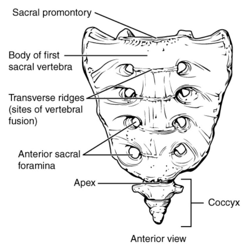
Sacral promontory: The sacral promontory is the prominent anterior projection at the upper end of the sacrum, formed by the body of the first sacral vertebra. It serves as a landmark for pelvic measurements and influences the alignment of the uterus and other pelvic organs.
Body of first sacral vertebra: The body of the first sacral vertebra is the largest and most robust segment of the sacrum, articulating with the fifth lumbar vertebra above. This structure bears significant weight and contributes to the sacrum’s overall stability.
Transverse ridges (sites of vertebral fusion): The transverse ridges are horizontal lines on the anterior sacrum, marking the fusion sites of the five sacral vertebrae during development. These ridges indicate where the individual vertebrae have fused into a single bone, enhancing its strength.
Anterior sacral foramina: The anterior sacral foramina are a series of openings on the front of the sacrum that allow the passage of the anterior rami of the sacral spinal nerves and blood vessels. These foramina are symmetrically arranged and play a crucial role in innervating the lower body.
Apex: The apex is the pointed, inferior end of the sacrum where it articulates with the coccyx, forming a flexible joint. This small structure supports the attachment of ligaments and aids in weight distribution to the coccyx.
Coccyx: The coccyx, or tailbone, is a small triangular bone formed by the fusion of four coccygeal vertebrae, located at the base of the sacrum. It provides attachment points for pelvic floor muscles and ligaments, supporting posture and aiding in childbirth.
Detailed Anatomical Overview
Exploring the Sacrum’s Anterior Structure
The anterior view reveals the sacrum’s unique triangular shape, a result of the fusion of five sacral vertebrae. This fusion creates a solid foundation that supports the spine and transfers weight to the pelvis through the sacroiliac joints. The sacral promontory acts as a critical reference point in obstetrics, influencing the size of the pelvic inlet.
- The body of first sacral vertebra is the strongest part, designed to handle compressive forces from the upper body.
- The transverse ridges highlight the developmental process, showing where growth plates once existed.
- The anterior sacral foramina allow the sacral nerves to exit, controlling functions like bladder and bowel movements.
Functional Anatomy and Clinical Importance
The sacrum’s anterior surface is concave, enhancing its weight-bearing capacity and protecting the spinal nerves within. The apex connects to the coccyx, allowing slight mobility that absorbs shock during sitting. This region’s anatomy is essential for diagnosing conditions like sacral fractures or nerve entrapments.
- The sacral promontory can be palpated to assess pelvic alignment or during labor to evaluate the birth canal.
- The body of first sacral vertebra may show signs of stress fractures in athletes due to repetitive loading.
- The transverse ridges serve as landmarks for surgical approaches to the sacrum.
Role of Foramina and Coccyx
The anterior sacral foramina are paired openings that correspond to the posterior foramina, forming a network for nerve and vascular passage. These foramina are vital for procedures like nerve blocks to manage lower back pain. The coccyx supports the pelvic diaphragm, including muscles like the levator ani, which are crucial for continence.
- The anterior sacral foramina provide pathways for the S1-S4 nerve roots, influencing lower limb sensation and movement.
- The apex allows the coccyx to move slightly, reducing pressure on the sacrum during prolonged sitting.
- The coccyx can be injured from falls, leading to coccydynia, a condition requiring careful management.
Clinical Applications and Anatomical Insights
Understanding the anterior sacrum is key for procedures like sacral colpopexy or epidural anesthesia. The sacral promontory is a guide for placing instruments during pelvic surgeries, ensuring precision. The body of first sacral vertebra’s alignment affects spinal curvature, potentially contributing to conditions like scoliosis.
- The transverse ridges help identify the level of vertebral fusion in imaging studies.
- The anterior sacral foramina are accessed in pain management to target specific nerve roots.
- The apex and coccyx are evaluated in cases of trauma to assess joint integrity.
The sacrum and coccyx, though compact, are integral to the skeletal framework, supporting posture and protecting neural structures. Their anterior view highlights the fusion process and nerve pathways, offering insights into both normal anatomy and clinical interventions. Mastery of this region enhances the ability to address related health issues with confidence and precision.
Conclusion
The anterior view of the sacrum and coccyx unveils a complex structure that underpins the body’s stability and mobility. This anatomical region’s role in weight distribution and nerve function underscores its importance in daily activities and medical practice. Exploring its labeled parts provides a deeper appreciation of its contributions to overall health and effective patient care.

