The cervical vertebrae form the upper portion of the spinal column, providing support and flexibility to the neck while protecting vital structures. This article delves into the anatomy of a typical cervical vertebra, exploring its key components and their roles in maintaining spinal health and movement.
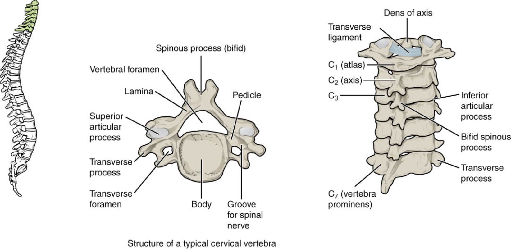
Vertebral foramen: The vertebral foramen is the large central opening in the cervical vertebra that houses the spinal cord, safeguarding it from external pressure. It allows for the passage of nerves that connect the brain to the body, ensuring proper neurological function.
Spinous process (bifid): The spinous process, uniquely bifid in cervical vertebrae, extends posteriorly and provides attachment points for muscles and ligaments. This forked structure enhances neck mobility and serves as a landmark for clinical assessments.
Lamina: The lamina forms the posterior part of the vertebral arch, connecting the pedicle to the spinous process and enclosing the vertebral foramen. It protects the spinal cord and contributes to the vertebra’s structural stability.
Pedicle: The pedicle is a short, sturdy column of bone that connects the vertebral body to the lamina, forming part of the vertebral arch. It supports the weight of the head and neck while anchoring the spinal column.
Superior articular process: The superior articular process projects upward to articulate with the vertebra above, facilitating smooth movement between cervical segments. It plays a key role in maintaining the alignment and flexibility of the neck.
Transverse process: The transverse process extends laterally from the vertebra, serving as an attachment site for muscles and ligaments that support neck movement. It also contains the transverse foramen, a critical feature of cervical anatomy.
Transverse foramen: The transverse foramen, located within the transverse process, allows the passage of the vertebral artery and vein, supplying blood to the brain. This structure is essential for cerebral circulation and distinguishes cervical vertebrae.
Body: The body is the anterior, weight-bearing portion of the vertebra, designed to absorb compressive forces from the head and neck. Its small size in cervical vertebrae reflects the region’s focus on flexibility rather than load-bearing.
Groove for spinal nerve: The groove for spinal nerve, located on the pedicle, provides a pathway for spinal nerves to exit the vertebral column. It ensures effective communication between the spinal cord and the rest of the body.
Dens of axis: The dens of axis, a projection from the second cervical vertebra (C2), fits into the atlas (C1) to enable head rotation. It is a unique feature visible in the context of cervical anatomy, particularly at the C1-C2 junction.
Transverse ligament: The transverse ligament holds the dens of the axis in place within the atlas, stabilizing the atlantoaxial joint. It prevents excessive movement, protecting the spinal cord from potential compression.
Inferior articular process: The inferior articular process extends downward to articulate with the vertebra below, ensuring continuity and stability in the cervical spine. It helps distribute mechanical loads across the spinal column.
Bifid spinous process: The bifid spinous process, a split posterior projection, provides additional attachment sites for neck muscles and ligaments. Its unique shape in cervical vertebrae enhances flexibility and range of motion.
C1 (atlas): The atlas, the first cervical vertebra, supports the skull and allows nodding movements through its articulation with the occipital condyles. It lacks a body and spinous process, distinguishing it from typical cervical vertebrae.
C2 (axis): The axis, the second cervical vertebra, features the dens, which serves as a pivot for head rotation. It works closely with the atlas to facilitate a wide range of neck movements.
C3: The third cervical vertebra represents a typical cervical structure, with a bifid spinous process and transverse foramina. It contributes to the neck’s flexibility and supports the spinal cord.
C7 (vertebra prominens): The seventh cervical vertebra, known as the vertebra prominens, has a long, non-bifid spinous process that is palpable under the skin. It marks the transition to the thoracic spine and serves as a clinical landmark.
Anatomical Structure and Function
The cervical vertebrae are designed for both support and mobility. The vertebral foramen protects the spinal cord, a critical neural pathway.
- The spinous process (bifid) enhances muscle attachment, aiding in neck flexion.
- The lamina and pedicle form a protective arch around the spinal cord.
- The superior articular process ensures smooth articulation with the vertebra above.
- The transverse process and transverse foramen support vascular and muscular functions.
This structure allows the neck to move freely while maintaining stability.
Ligaments and Joint Stability
Ligaments play a key role in stabilizing the cervical spine. The transverse ligament secures the dens of axis, ensuring joint integrity.
- The inferior articular process connects with the vertebra below, distributing loads.
- The bifid spinous process provides leverage for muscle action.
- The groove for spinal nerve facilitates nerve exit, supporting sensory and motor functions.
- The C1 (atlas) and C2 (axis) form a unique joint, enabling head rotation.
These elements prevent excessive motion and protect the spinal cord.
Clinical Relevance and Movement
The cervical vertebrae’s anatomy has significant clinical importance. The transverse foramen’s vascular role is critical in cases of arterial injury.
- The superior articular process and inferior articular process are key in diagnosing joint disorders.
- The C7 (vertebra prominens) serves as a landmark for spinal assessments.
- The body absorbs compressive forces, influencing posture and alignment.
- The dens of axis is a focus in upper cervical injuries like dislocations.
Understanding these features aids in effective diagnosis and treatment.
The cervical vertebrae, with their intricate vertebral foramen and supportive spinous process (bifid), form a flexible yet protective spinal segment. The transverse foramen, pedicle, and lamina ensure vascular and neural integrity, while the C1 (atlas) and C2 (axis) enable unique head movements. The C7 (vertebra prominens) marks a transition point, emphasizing the region’s role in overall spinal health and mobility.

