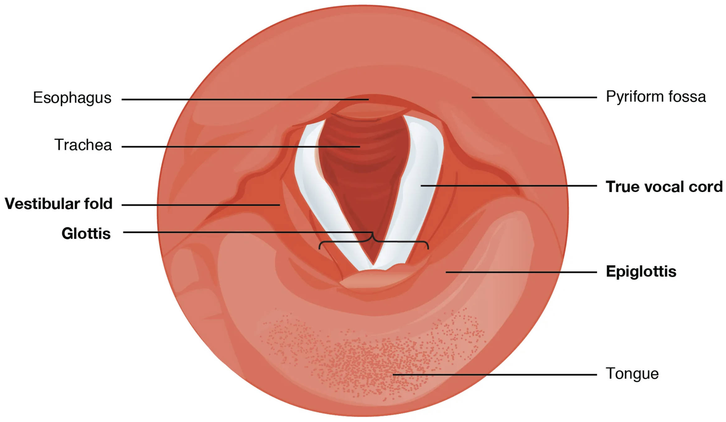
The vocal cords, a central feature of the larynx, are critical for voice production and airway protection, viewed here from the laryngopharynx looking downward. Comprising the true vocal cords and vestibular folds, this region facilitates phonation through intricate muscle and nerve coordination while safeguarding the trachea during swallowing. A superior view of these structures provides a unique perspective on their arrangement and function, enhancing comprehension of their physiological significance.
Key Anatomical Labels in the Diagram
This section delves into each labeled component, offering insights into their roles and positions within the laryngeal framework.
Esophagus: The esophagus is the muscular tube posterior to the larynx, transporting food from the pharynx to the stomach via peristalsis. It remains closed during breathing to prevent air entry into the digestive tract.
Trachea: The trachea, or windpipe, extends from the larynx to the bronchi, conducting air to the lungs with support from C-shaped cartilages. It ensures a patent airway beneath the vocal cords.
Vestibular fold: The vestibular fold, or false vocal cord, lies above the true vocal cord, protecting the airway during swallowing. It contributes minimally to sound production but aids in closing the larynx.
Glottis: The glottis is the opening between the vocal cords, serving as the primary passageway for air during respiration and phonation. Its size adjusts through muscle action, influencing pitch and volume.
Pyriform fossa: The pyriform fossa is a paired recess on either side of the larynx, channeling food into the esophagus during swallowing. It also assists in directing airflow and protecting the airway.
True vocal cord: The true vocal cord vibrates to produce sound as air passes, controlled by the recurrent laryngeal nerve. It is essential for speech, coughing, and maintaining airway pressure.
Epiglottis: The epiglottis is a cartilage flap that covers the larynx during swallowing, preventing food from entering the trachea. It lifts during breathing to allow air passage into the lungs.
Tongue: The tongue occupies the oral cavity floor, aiding in food manipulation and taste perception. It helps propel food toward the pharynx, coordinating with the epiglottis during swallowing.
The Vocal Cord Framework
The vocal cords and surrounding structures form a dynamic system within the larynx. This setup supports both respiratory and phonatory functions.
- The true vocal cords are anchored by the thyroid and arytenoid cartilages.
- Vestibular folds provide an additional layer of airway protection.
- The glottis adjusts dynamically, narrowing for phonation or widening for breathing.
- The epiglottis and pyriform fossa work together to guide food safely.
- Tracheal support ensures continuous airflow to the lungs.
Functional Roles in Phonation and Respiration
The vocal cords play a dual role in sound production and airway management. Their anatomy enables precise control over these processes.
- The true vocal cords vibrate to generate pitch, modulated by muscle tension.
- The glottis opens widely during respiration, allowing efficient air exchange.
- Vestibular folds close to protect the airway during swallowing.
- The epiglottis prevents aspiration, coordinating with the tongue.
- The trachea maintains a clear path for oxygenated air.
Supporting Structures and Their Contributions
Surrounding anatomical features enhance the vocal cords’ effectiveness. These elements ensure seamless integration with the broader airway system.
- The pyriform fossa directs food, reducing the risk of choking.
- The esophagus remains passive during breathing, activating during swallowing.
- The tongue propels food, supporting the epiglottis’s protective role.
- The vestibular folds cushion the true vocal cords from trauma.
- These structures adapt to pressure changes, maintaining functionality.
Clinical Relevance and Anatomical Variations
Understanding vocal cord anatomy is essential for diagnosing and treating related conditions. Variations can influence clinical approaches.
- Vocal cord paralysis, often due to recurrent nerve damage, affects voice.
- Epiglottitis can obstruct the airway, requiring urgent medical attention.
- Glottal stenosis may result from scarring, impacting breathing.
- Pyriform fossa tumors can interfere with swallowing and speech.
- Laryngoscopy helps visualize these structures for accurate diagnosis.
The vocal cords, viewed superiorly, reveal a sophisticated interplay of cartilage, muscle, and nerve that supports voice and respiration. By studying this anatomical layout, one gains a deeper appreciation for the larynx’s role in human communication and survival, showcasing the elegance of its design.

