Understanding the Divisions of the Pharynx: A Detailed Anatomical Guide
The pharynx is a crucial passageway in the human body, serving as a shared conduit for air and food between the nasal cavity and the larynx or esophagus. This muscular tube, divided into three distinct regions—nasopharynx, oropharynx, and laryngopharynx—plays an essential role in respiration, swallowing, and even speech production. Exploring its anatomical divisions through detailed diagrams offers a clearer perspective on its structure and the functions each segment supports.
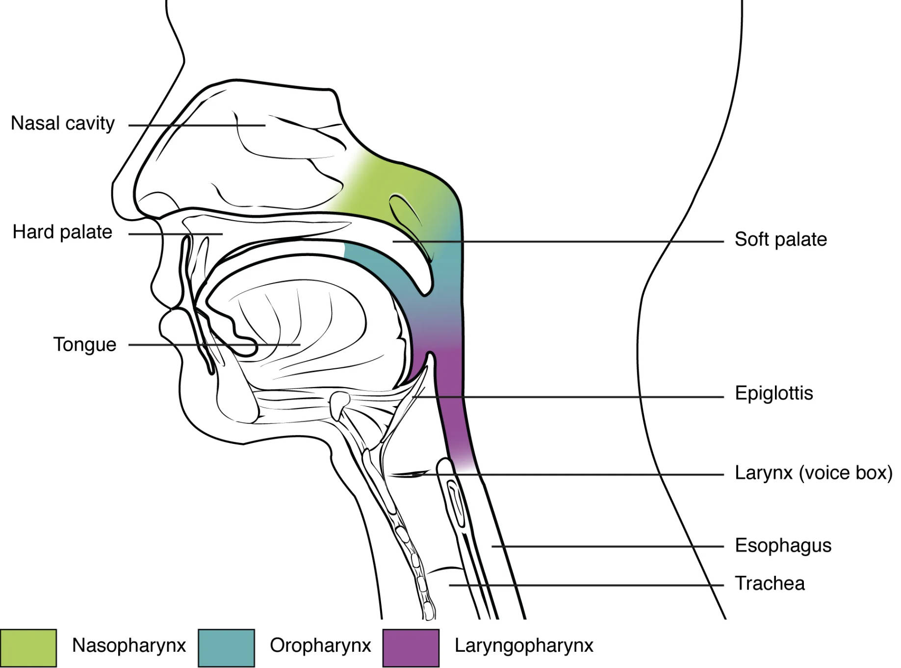
Key Anatomical Labels in the Diagram
This section provides an overview of each labeled component, highlighting their specific roles and locations within the pharyngeal structure.
Nasal cavity: The nasal cavity is the uppermost air passage, filtering and humidifying air before it reaches the pharynx. It connects to the nasopharynx, serving as the initial entry point for respiration and olfaction.
Hard palate: The hard palate forms the bony anterior roof of the mouth, separating the oral cavity from the nasal cavity. It provides a stable platform for chewing and aids in directing airflow into the nasopharynx.
Tongue: The tongue occupies the floor of the oral cavity, assisting in food manipulation and taste perception. It helps propel food into the oropharynx during the swallowing process.
Soft palate: The soft palate is a muscular extension behind the hard palate, elevating to close off the nasopharynx during swallowing. It also plays a key role in modulating airflow for speech.
Epiglottis: The epiglottis is a cartilage flap at the base of the tongue, closing the larynx during swallowing to prevent food entry into the airway. It reopens to allow air passage, coordinating respiration and digestion.
Larynx (voice box): The larynx, located below the pharynx, houses the vocal cords and produces sound through vocal fold vibration. It connects to the trachea, ensuring air reaches the lungs while supporting phonation.
Esophagus: The esophagus is the muscular tube that transports food from the laryngopharynx to the stomach via peristalsis. It remains closed during breathing to prevent air entry into the digestive tract.
Trachea: The trachea, or windpipe, extends from the larynx to the bronchi, conducting air to the lungs. It is reinforced with cartilage rings to maintain an open airway.
Nasopharynx: The nasopharynx is the uppermost pharyngeal region, connecting the nasal cavity to the oropharynx and facilitating air passage. It contains the pharyngeal tonsil, which aids in immune defense.
Oropharynx: The oropharynx lies between the soft palate and the epiglottis, serving as a common pathway for air and food. It houses the palatine tonsils, contributing to immune surveillance.
Laryngopharynx: The laryngopharynx, or hypopharynx, is the lowest pharyngeal section, transitioning to the esophagus and larynx. It directs food to the stomach and air to the trachea during swallowing and breathing.
The Upper Pharyngeal Region: Nasopharynx
The nasopharynx sets the stage for respiratory function with its strategic location. This area supports continuous airflow and immune protection.
- The nasopharynx remains open during breathing, allowing air to flow from the nasal cavity.
- The pharyngeal tonsil within this region filters pathogens, reducing respiratory infections.
- It connects to the auditory tube, equalizing middle ear pressure.
- Mucous membranes humidify air, protecting the delicate respiratory lining.
- Structural deviations here can lead to chronic sinus issues or sleep apnea.
The Middle Pharyngeal Region: Oropharynx
The oropharynx bridges the oral cavity and lower pharynx, handling both digestion and respiration. Its design supports a dual-purpose function seamlessly.
- The oropharynx receives food propelled by the tongue during swallowing.
- Palatine tonsils here provide a first line of defense against ingested pathogens.
- It collaborates with the soft palate to prevent nasal reflux during eating.
- The epiglottis guards the airway entrance, ensuring safe passage of food.
- Inflammation in this area often signals tonsillitis or pharyngitis.
The Lower Pharyngeal Region: Laryngopharynx
The laryngopharynx serves as a critical junction for air and food pathways. Its anatomy ensures efficient separation of respiratory and digestive routes.
- The laryngopharynx directs food into the esophagus while air moves to the larynx.
- It is supported by muscles like the inferior constrictor, aiding swallowing.
- The epiglottis and vocal cords coordinate to protect the airway.
- This region is prone to reflux, where stomach acid can irritate the tissues.
- Surgical interventions here may address swallowing difficulties or tumors.
Functional Roles of the Pharyngeal Divisions
Each pharyngeal division contributes uniquely to overall physiology. Their coordinated action is vital for survival.
- The nasopharynx filters and warms air, enhancing lung health.
- The oropharynx supports speech by modulating airflow with the tongue.
- The laryngopharynx ensures food and air follow distinct paths.
- Vocal cord vibration in the larynx produces a range of pitches.
- These regions adapt to pressure changes, maintaining efficient function.
Clinical Significance and Anatomical Variations
Understanding pharyngeal anatomy aids in diagnosing and treating related conditions. Variations can impact treatment strategies significantly.
- Nasopharyngeal obstruction may result from adenoid hypertrophy, affecting breathing.
- Oropharyngeal cancers often originate in the tonsillar tissue, requiring early detection.
- Laryngopharyngeal reflux can damage the larynx, leading to hoarseness.
- Tracheal compression from external masses may necessitate imaging studies.
- Anatomical knowledge guides procedures like laryngoscopy for diagnosis.
The pharynx’s division into nasopharynx, oropharynx, and laryngopharynx reflects its versatile role in respiration, digestion, and communication. By studying these regions through detailed anatomical diagrams, one gains a deeper appreciation for their interconnected functions, underscoring the complexity and resilience of the human airway system.

