The heart’s efficient pumping action relies on the precise movement of blood through its chambers, and this diagram illustrates the critical pathway from the left atrium to the left ventricle. Featuring transverse and frontal sections with the atria and vessels removed, the image highlights the mitral valve’s role in facilitating this flow while the aortic semilunar valve prevents backflow. Examining this diagram provides a deeper understanding of the heart’s structure and the mechanisms that ensure oxygenated blood reaches the body effectively.
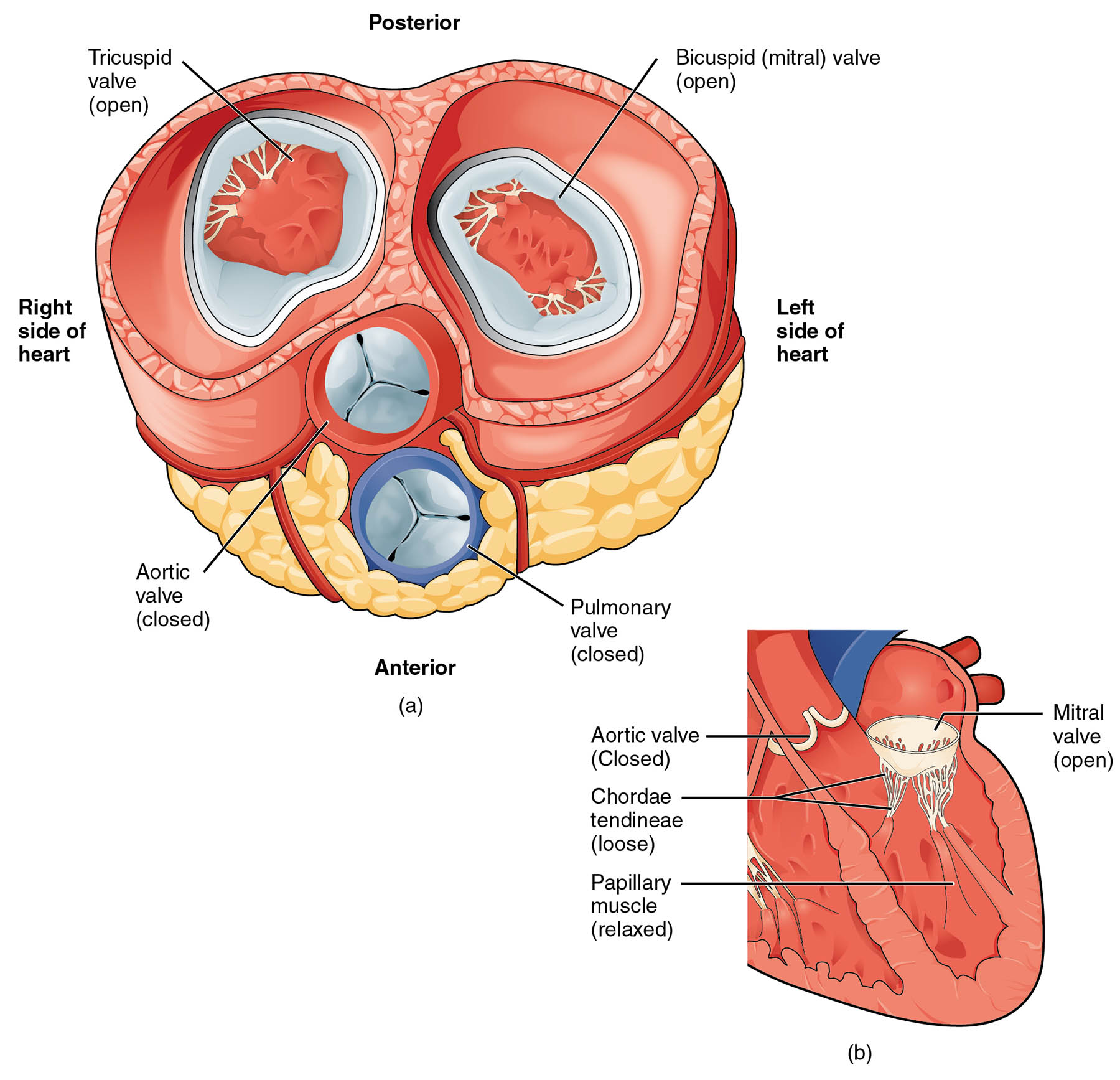
Mitral valve: The mitral valve, or bicuspid valve, separates the left atrium from the left ventricle, opening to allow oxygenated blood to flow into the ventricle during diastole. It closes during systole to prevent backflow, supported by chordae tendineae and papillary muscles for stability.
Left ventricle: The left ventricle is the thick-walled chamber that pumps oxygenated blood into the aorta for systemic circulation, generating high pressure to meet the body’s demands. Its robust structure is designed to handle the force required to propel blood throughout the body.
Aortic semilunar valve: The aortic semilunar valve, located between the left ventricle and aorta, consists of three cusps that open to release blood during systole and close to prevent backflow from the aorta. This valve ensures that blood moves forward into the systemic circulation without returning to the ventricle.
Anatomical Structure of Blood Flow Pathway
The heart’s left side is responsible for distributing oxygenated blood, and this diagram showcases its key components. The detailed sections reveal how blood transitions through the chambers and valves.
- The mitral valve serves as a gateway, opening to fill the left ventricle with blood from the lungs.
- The left ventricle’s muscular walls are tailored for forceful contraction, driving blood onward.
- The aortic semilunar valve acts as a one-way door, protecting the ventricle from pressure reversal.
- This pathway ensures efficient delivery of oxygen-rich blood to the aorta and beyond.
The removal of atria and vessels focuses attention on these critical structures.
Functional Roles in Circulation
The valves and chambers work together to maintain unidirectional blood flow, as depicted in this image. Their synchronized action is vital for effective cardiac performance.
- The mitral valve opens during relaxation, allowing blood to fill the left ventricle efficiently.
- The left ventricle contracts to eject blood, relying on the aortic semilunar valve to open.
- The aortic semilunar valve closes post-ejection, preventing blood from re-entering the ventricle.
- This cycle supports the heart’s role as a high-pressure pump for systemic circulation.
Any disruption can lead to compromised oxygen delivery.
Physical Characteristics and Clinical Relevance
The physical properties of these structures reflect their specialized functions within the heart. Their design supports the high demands of left-sided circulation.
- The mitral valve’s two cusps are reinforced to withstand the pressure of left atrial filling.
- The left ventricle’s thick myocardium enables it to generate significant force for systemic flow.
- The aortic semilunar valve’s elastic cusps adapt to rapid pressure changes during the cardiac cycle.
- Conditions like mitral regurgitation or aortic stenosis can arise from valve or ventricular issues.
Echocardiography helps assess these structures for abnormalities.
Importance in Cardiac Health
Maintaining the integrity of the mitral valve, left ventricle, and aortic semilunar valve is crucial for long-term heart function. Their health influences overall circulatory efficiency.
- A functional mitral valve prevents leakage, ensuring proper ventricular filling.
- The left ventricle’s strength supports sustained output, vital for physical activity.
- The aortic semilunar valve’s competence avoids backpressure, protecting heart muscle.
- Surgical interventions, like valve replacement, may be needed if degeneration occurs.
Regular monitoring through clinical exams supports early detection of issues.
Conclusion
This diagram of blood flow from the left atrium to the left ventricle offers a detailed view of the mitral valve, left ventricle, and aortic semilunar valve, illustrating their roles in oxygenated blood circulation. By highlighting the valve dynamics and chamber function, it emphasizes the heart’s precision in maintaining systemic flow. This understanding enhances appreciation for cardiac anatomy and the importance of preserving these structures for optimal health.

