Coarctation of the aorta is a congenital heart defect characterized by an abnormal narrowing of the aorta, which restricts blood flow to the lower body. This diagram illustrates the anatomical location and impact of this narrowing, offering a clear visual representation of how it affects the heart and circulatory system. Exploring this image provides essential insights into the condition’s implications and the importance of timely diagnosis and treatment.
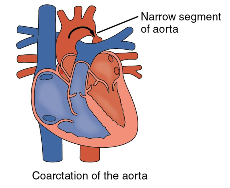
Aortic arch: The aortic arch is the curved portion of the aorta that carries oxygenated blood from the left ventricle to the descending aorta, distributing it to the upper body. In coarctation, a narrowing in this region increases resistance, forcing the heart to work harder to pump blood.
Narrowed segment: The narrowed segment is the constricted area of the aorta, typically located near the ductus arteriosus, which limits blood flow to the lower body and extremities. This stenosis can lead to hypertension in the upper body and weak pulses in the lower limbs.
Descending aorta: The descending aorta carries blood from the aortic arch to the abdominal aorta and lower body organs, supplying oxygen-rich blood to tissues. A coarctation upstream reduces pressure and flow in this section, potentially causing organ malperfusion.
Left ventricle: The left ventricle pumps oxygenated blood into the aorta, generating the pressure needed for systemic circulation. In coarctation of the aorta, this chamber may hypertrophy due to the increased workload from the obstruction.
Anatomical Overview of Coarctation of the Aorta
The aorta, the body’s main artery, plays a central role in distributing oxygenated blood, and this diagram highlights its normal and affected states. Understanding the anatomy aids in recognizing the defect’s impact on circulation.
- The aortic arch serves as the initial pathway for blood leaving the heart, branching to major arteries.
- The narrowed segment creates a bottleneck, altering blood pressure dynamics across the body.
- The descending aorta relies on adequate flow from the arch, which is compromised by the constriction.
- The left ventricle adapts by thickening its walls, a response to the increased afterload.
This defect often presents with collateral circulation development as a compensatory mechanism.
Physiological Impact and Symptoms
Coarctation of the aorta disrupts normal blood flow, leading to a range of physiological effects. The diagram illustrates how this narrowing influences cardiovascular function.
- The aortic arch experiences elevated pressure proximal to the narrowed segment, causing upper body hypertension.
- Reduced flow through the descending aorta can result in cold feet, leg cramps, or delayed growth in children.
- The left ventricle’s hypertrophy increases the risk of heart failure if the obstruction persists.
- Symptoms like headaches or nosebleeds may arise due to high pressure in the upper circulation.
Echocardiography or MRI can confirm the location and severity of the narrowing.
Causes and Risk Factors
The development of coarctation of the aorta involves genetic and environmental factors during fetal development. Identifying these helps in understanding its origins.
- Genetic syndromes, such as Turner syndrome, are associated with a higher incidence of this defect.
- Maternal factors like hypertension or diabetes during pregnancy may contribute to congenital anomalies.
- Abnormalities in the ductus arteriosus closure process can lead to the narrowed segment formation.
- Prenatal ultrasound screening can detect aortic narrowing, enabling early planning.
Folic acid intake during pregnancy may reduce the risk of related congenital defects.
Diagnosis and Treatment Options
Diagnosing and managing coarctation of the aorta requires a comprehensive approach based on the defect’s severity. Advanced imaging is key to assessing the condition.
- Doppler echocardiography measures the pressure gradient across the narrowed segment to evaluate obstruction.
- Asymptomatic cases may be monitored, but intervention is often needed to prevent complications.
- Balloon angioplasty or surgical repair widens the aortic arch, restoring normal flow to the descending aorta.
- Medications like beta-blockers manage hypertension until definitive treatment is performed.
Long-term follow-up is essential to monitor for restenosis or aneurysm formation.
Clinical Relevance and Long-Term Outlook
Understanding the implications of coarctation of the aorta is crucial for long-term health management. The defect’s effects vary depending on its severity and treatment.
- The aortic arch’s pressure changes can lead to early coronary artery disease if untreated.
- Adequate blood supply to the descending aorta is vital for lower body organ function, including the kidneys.
- The left ventricle’s health improves post-repair, reducing heart failure risk.
- Lifelong monitoring ensures the narrowed segment does not recur, with regular blood pressure checks.
Lifestyle modifications, such as a heart-healthy diet, support overall cardiovascular wellness.
Conclusion
This diagram of coarctation of the aorta provides a detailed illustration of the aortic arch, narrowed segment, descending aorta, and left ventricle, showcasing the impact of this congenital defect on blood flow. By highlighting the narrowing’s effect on circulation and heart function, it emphasizes the need for early detection and intervention. This knowledge empowers individuals to address the challenges of coarctation effectively, improving outcomes with appropriate care.

