The palatine tonsil, located on either side of the throat, serves as a first line of defense in the immune system by trapping pathogens entering through the mouth and nose. This histological image, captured at a magnification of ×40, reveals the intricate cellular structure of the palatine tonsil, highlighting its role in immune surveillance. Exploring this micrograph provides a deeper understanding of its anatomical features and functional significance in maintaining health.
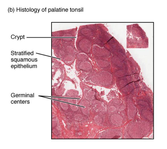
Crypt: The crypt is a deep invagination in the tonsil’s surface that increases its exposure to pathogens, allowing for effective trapping and processing. It contains lymphoid tissue and is lined with stratified squamous epithelium, enhancing its immune function.
Stratified squamous epithelium: The stratified squamous epithelium forms a protective outer layer over the tonsil, shielding underlying tissues from mechanical damage and microbial invasion. This multi-layered structure also supports the crypts, aiding in antigen capture and immune activation.
Germinal centers: Germinal centers are regions within the tonsil’s lymphoid follicles where B lymphocytes proliferate and differentiate into plasma cells, producing antibodies. These areas are critical for mounting a robust immune response against inhaled or ingested pathogens.
Anatomical Structure of the Palatine Tonsil
The palatine tonsil is a mass of lymphoid tissue situated at the back of the oral cavity, typically measuring about 2-3 cm in length. Its histological makeup is designed to optimize immune protection in the upper respiratory tract.
- The crypt extends deep into the tonsil, creating a large surface area for pathogen interaction.
- These invaginations are lined with the stratified squamous epithelium, which provides a durable barrier against physical and microbial threats.
- The germinal centers within the lymphoid follicles are bustling with immune activity, supporting B-cell maturation.
- This structure collectively enhances the tonsil’s ability to filter and respond to antigens, playing a key role in local immunity.
The tonsil’s position and architecture make it an essential component of the Waldeyer’s ring, a ring of lymphoid tissue encircling the pharynx.
Microscopic Features and Functionality
This histological image, magnified at ×40, showcases the tonsil’s cellular and structural details. The clarity of the micrograph aids in understanding its physiological roles.
- The crypt’s irregular shape traps debris and microorganisms, initiating an immune response at the entry point.
- The stratified squamous epithelium’s multiple layers offer resilience, adapting to the constant exposure of the oral cavity.
- Within the lymphoid tissue, germinal centers are sites of intense B-cell proliferation, crucial for antibody production.
- The interplay of these components ensures efficient pathogen detection and neutralization, supporting overall immune health.
The tonsil’s ability to regenerate lymphoid tissue further underscores its dynamic role in immunity.
Physical Characteristics and Clinical Relevance
The palatine tonsil’s physical properties reflect its function as an immune sentinel. It varies in size with age and can become enlarged during infections, a condition known as tonsillitis.
- The crypt’s depth allows it to harbor a diverse microbial community, which can sometimes lead to inflammation if unchecked.
- The stratified squamous epithelium’s thickness protects against abrasions from food or breathing, maintaining tissue integrity.
- The germinal centers’ activity increases during infections, producing antibodies like IgA to combat pathogens.
- Chronic enlargement of the tonsils may require medical evaluation, as it can affect breathing or swallowing.
Understanding these features helps in assessing tonsil-related conditions and their impact on health.
Conclusion
The histological image of the palatine tonsil offers a detailed look at its critical role in immune defense within the upper respiratory tract. The crypt, stratified squamous epithelium, and germinal centers work synergistically to trap, process, and neutralize pathogens, safeguarding the body. This exploration of the tonsil’s microstructure provides a solid foundation for studying its functions and addressing related health concerns effectively.

