The spleen, a vital organ in the human body, plays a crucial role in filtering blood and supporting the immune system. This magnified histological image provides a detailed view of the spleen’s intricate structure, highlighting key components essential for its function. By examining this micrograph, one can gain a deeper understanding of how the spleen processes antigens and maintains blood quality, making it an invaluable resource for studying human anatomy and physiology.
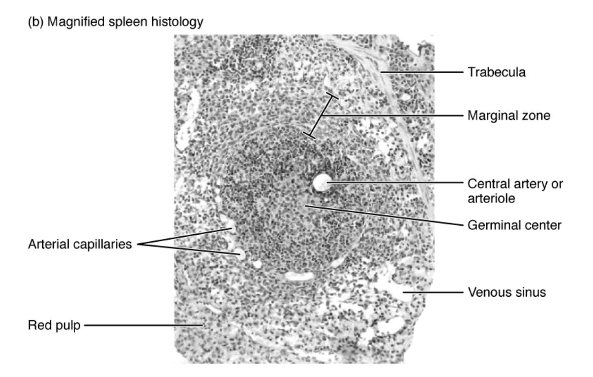
Trabecula: The trabecula consists of connective tissue that provides structural support to the spleen, forming a framework for its soft tissue. It helps anchor the organ and allows for the passage of blood vessels and nerves, ensuring proper circulation and innervation.
Marginal zone: The marginal zone serves as a transitional area between the red pulp and white pulp, acting as a filter for blood-borne pathogens and antigens. It contains specialized macrophages and dendritic cells that capture and present antigens to lymphocytes, initiating an immune response.
Central artery or arteriole: The central artery or arteriole is a key blood vessel that supplies oxygenated blood to the white pulp, facilitating immune cell activity. It branches into smaller capillaries, distributing blood to support the spleen’s filtration and immune functions.
Germinal center: The germinal center is a site within the white pulp where B lymphocytes proliferate and mature, producing antibodies to combat infections. It is a dynamic region where immune responses are fine-tuned, enhancing the body’s defense mechanisms.
Venous sinus: The venous sinus collects filtered blood from the red pulp, allowing deoxygenated blood to exit the spleen and return to circulation. These sinuses are lined with endothelial cells that regulate blood flow and facilitate the removal of damaged cells.
Arterial capillaries: Arterial capillaries extend from the central artery, delivering blood to the spleen’s functional areas for filtration and immune processing. They play a critical role in distributing nutrients and oxygen while enabling the spleen to monitor and cleanse the blood.
Red pulp: The red pulp is rich in venous sinuses and macrophages, primarily responsible for filtering old or damaged red blood cells from circulation. It also serves as a reservoir for blood, releasing it during times of need, such as hemorrhage.
Detailed Anatomical Overview of the Spleen
The spleen is an organ located in the upper left quadrant of the abdomen, nestled between the stomach and diaphragm. It is composed of two main regions: the red pulp and the white pulp, each with distinct roles in maintaining blood quality and immunity.
- The red pulp is characterized by its reddish hue due to the presence of numerous red blood cells, making it a key player in hemocytoblasts removal.
- This area contains venous sinuses that trap and destroy old or abnormal erythrocytes, ensuring a healthy blood supply.
- The white pulp surrounds the central arteries and is densely populated with lymphocytes, forming the spleen’s immune hub.
- Here, the germinal center within the white pulp is where B cells undergo maturation, a process vital for producing specific antibodies.
The marginal zone acts as a critical interface, bridging the red and white pulp to facilitate antigen capture. This region is lined with macrophages that engulf pathogens, presenting them to lymphocytes for an immune response.
Microscopic Structure and Functionality
The histological image reveals the spleen’s complex microstructure under a magnification of ×660. This level of detail showcases the interplay between its vascular and lymphatic components.
- The trabecula provides a scaffold that supports the spleen’s soft tissue, allowing it to withstand physical stress.
- Blood enters through the central artery or arteriole, branching into arterial capillaries that nourish the tissue and support immune surveillance.
- The venous sinus then collects the filtered blood, ensuring efficient circulation and waste removal.
- This intricate network highlights the spleen’s dual role in filtration and immunity, with the germinal center being a focal point for lymphocyte activation.
Clinical Relevance and Physical Characteristics
Understanding the spleen’s anatomy is essential for recognizing its physiological significance. The organ weighs approximately 150-200 grams in adults and varies in size depending on individual health status.
- The red pulp‘s ability to store blood makes it a vital backup during blood loss, releasing reserves as needed.
- Its spongy texture, supported by the trabecula, allows it to expand and contract with blood volume changes.
- The marginal zone‘s role in antigen sequestration underscores its importance in preventing systemic infections.
- Damage to the venous sinus or arterial capillaries can impair blood flow, potentially leading to splenomegaly or other complications.
Conclusion
The magnified spleen histology image offers a window into the organ’s sophisticated structure and function, essential for blood filtration and immune defense. By studying the trabecula, marginal zone, central artery or arteriole, germinal center, venous sinus, arterial capillaries, and red pulp, one can appreciate the spleen’s critical role in maintaining health. This detailed exploration serves as a foundation for further research and clinical applications, emphasizing the organ’s importance in human physiology.

