The spleen, a key organ in the upper left abdomen, plays an essential role in filtering blood and supporting the immune system. Positioned near the stomach and protected by the diaphragm, it acts as a reservoir for blood cells and a site for lymphocyte maturation, contributing to overall health and defense against pathogens. This cross-sectional illustration provides a comprehensive look at its anatomy, highlighting the intricate network of structures that define its function.
Labeled Components of the Spleen
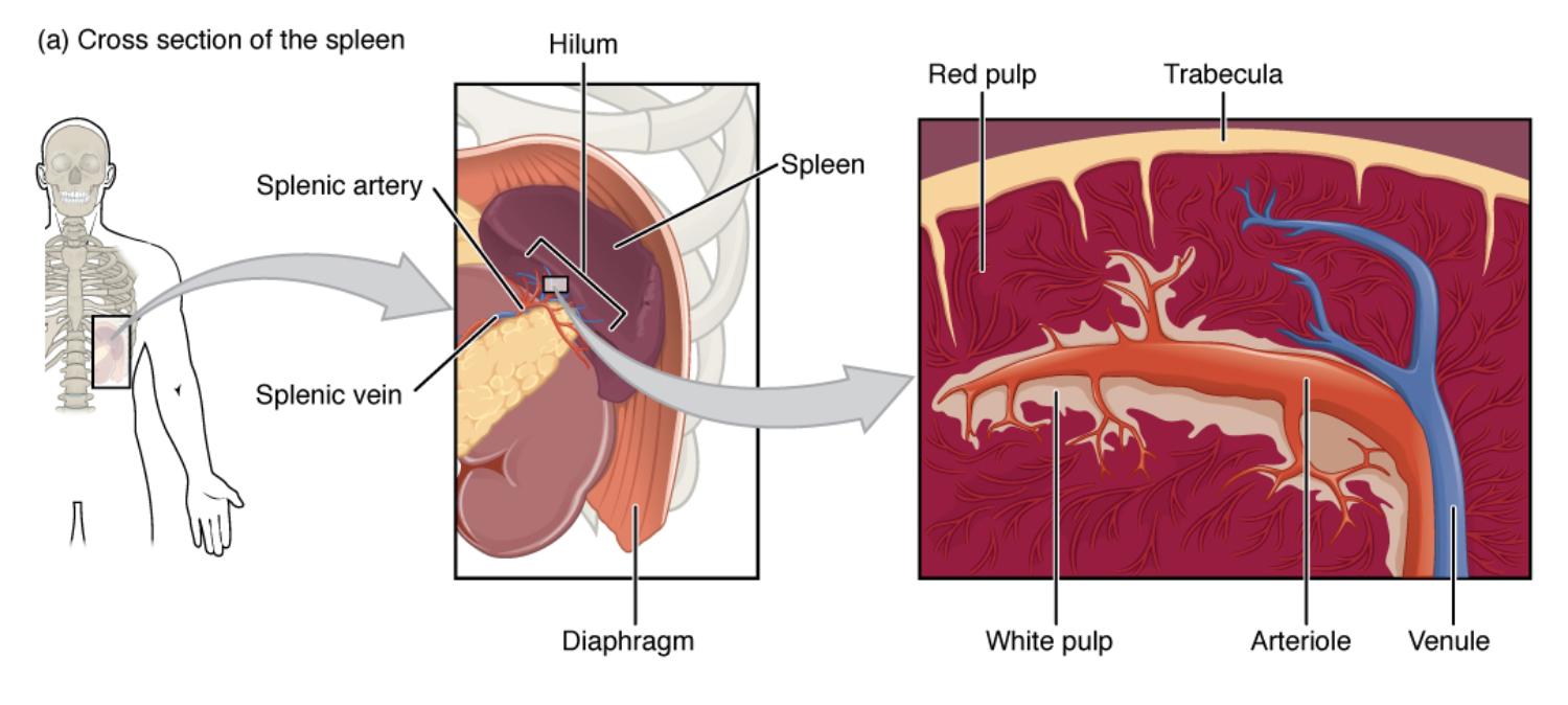
Hilum: This is the point where the splenic artery and vein enter and exit the spleen, serving as a critical junction for blood flow. It also accommodates lymphatic vessels and nerves, ensuring the organ’s integration with the circulatory system.
Red pulp: This region filters blood, removing old or damaged red blood cells and platelets with the help of macrophages. It contains a network of sinuses that process approximately 10% of the body’s blood volume daily.
Trabecula: These connective tissue extensions provide structural support, dividing the spleen into compartments. They guide blood vessels and nerves, maintaining the organ’s shape and function.
Spleen: The organ itself is a soft, vascular structure located beneath the diaphragm, adjacent to the stomach. It plays a dual role in blood filtration and immune response, adapting its size based on physiological demands.
Diaphragm: This muscular structure separates the thoracic and abdominal cavities, offering protection to the spleen. It also influences blood flow through respiratory movements, impacting splenic function.
White pulp: This lymphoid tissue surrounds the central arteries, hosting lymphocytes for immune activation. It is essential for initiating responses against blood-borne antigens.
Splenic artery: This major vessel delivers oxygenated blood to the spleen from the celiac trunk via the hilum. It branches into smaller arterioles within the organ to support its metabolic needs.
Splenic vein: This vessel drains deoxygenated blood from the spleen, returning it to the hepatic portal system. It exits through the hilum, completing the circulatory cycle.
Arteriole: These small arteries branch from the splenic artery, supplying blood to the white pulp and red pulp regions. They are vital for delivering oxygen to immune and filtration sites.
Venule: These small veins collect blood from the splenic sinuses, channeling it toward the splenic vein. They ensure the return of filtered blood to the systemic circulation.
Anatomical Structure of the Spleen
The spleen’s cross-sectional view reveals a sophisticated design tailored for its immune and hematological roles.
- The hilum serves as the entry and exit point for the splenic artery and vein, anchoring the organ’s vascular supply.
- Red pulp dominates with its sinus network, filtering out damaged cells and pathogens efficiently.
- Trabecula provide a supportive framework, preventing the spleen from collapsing under pressure.
- The diaphragm offers a protective barrier, while the spleen’s proximity to the stomach enhances its blood supply.
- White pulp forms lymphoid nodules around arterioles, where immune cells are activated.
- The splenic artery and vein ensure a continuous blood flow, with arterioles and venules distributing it within the organ.
This illustration underscores the spleen’s strategic location and vascular connectivity.
Physiological Functions of the Spleen
The spleen performs critical tasks, balancing blood filtration with immune defense.
- Red pulp removes senescent red blood cells, recycling iron for new hemoglobin production.
- White pulp initiates adaptive immunity, with lymphocytes responding to antigens in the blood.
- The hilum facilitates the entry of oxygenated blood via the splenic artery and the exit of deoxygenated blood through the splenic vein.
- Trabecula support the structural integrity, allowing efficient blood flow through the red pulp.
- Arterioles deliver blood to immune sites, while venules collect filtered blood for circulation.
- Macrophages within the red pulp also engulf bacteria, enhancing innate immunity.
This dual functionality highlights the spleen’s role in maintaining blood quality and immune readiness.
Clinical Relevance of Spleen Anatomy
Understanding the spleen’s structure aids in diagnosing and managing related health issues.
- An enlarged spleen, or splenomegaly, may indicate infections or hematologic disorders, often visible at the hilum.
- Red pulp dysfunction can lead to hemolytic anemia, reducing the spleen’s filtration capacity.
- White pulp abnormalities may weaken immune responses, increasing susceptibility to infections.
- The diaphragm’s protective role is critical in traumatic injuries affecting the spleen.
- Trabecula provide structural clues in cases of splenic rupture or chronic inflammation.
- The splenic artery and vein are key focus areas during splenectomy to control bleeding.
This anatomical knowledge is vital for effective clinical decision-making.
Developmental and Adaptive Features
The spleen adapts throughout life, reflecting its dynamic role in health.
- The hilum develops early, establishing vascular connections during fetal growth.
- Red pulp expands during infections, increasing filtration to handle higher blood loads.
- Trabecula strengthen with age, supporting the spleen’s increasing workload.
- The diaphragm’s movement influences splenic blood flow, adapting to respiratory changes.
- White pulp matures postnatally, enhancing immune competence over time.
- The splenic artery and vein adjust their caliber to meet the spleen’s varying demands.
This adaptability ensures the spleen remains functional across different life stages.
The spleen’s intricate anatomy, as shown in this cross-sectional view, underscores its vital role in blood filtration and immune support. Its strategic placement near the stomach and protection by the diaphragm make it a fascinating organ, offering valuable insights into human physiology and health for anyone eager to explore its functions.

