The spleen is a vital organ nestled in the upper left abdomen, playing a central role in filtering blood and supporting immune function. Connected to the stomach and protected by the diaphragm, it serves as a reservoir for blood cells and a site for lymphocyte activation, contributing to the body’s defense against infections. This detailed view, including a cross-section and magnified histology, offers a window into the spleen’s complex structure and its critical physiological roles.
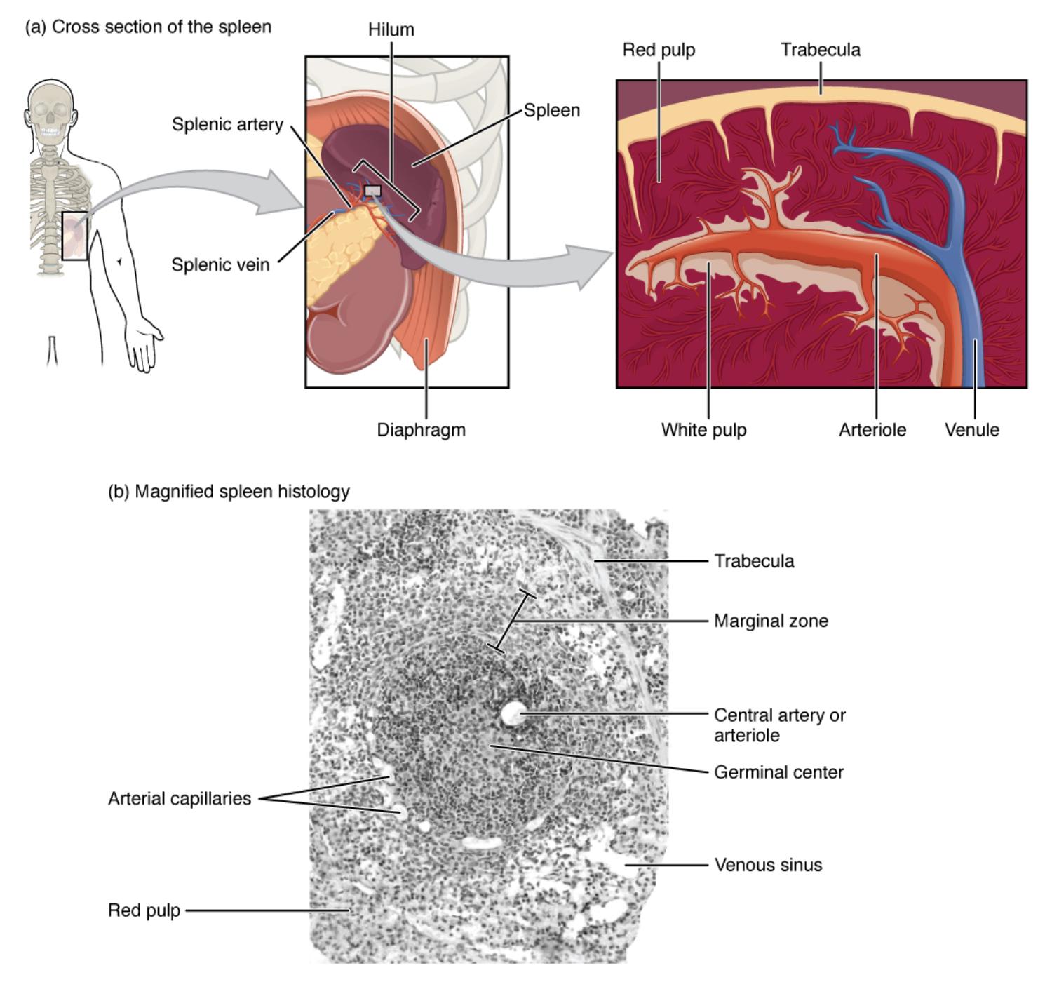
Labeled Components of the Spleen
Hilum: This is the entry and exit point for the splenic artery and vein, allowing blood to flow into and out of the spleen. It also serves as a pathway for nerves and lymphatic vessels, facilitating the organ’s connectivity.
Red pulp: Comprising a network of sinuses and cords, the red pulp filters blood, removing old or damaged red blood cells and platelets. It is rich in macrophages that phagocytose pathogens and cellular debris.
Trabecula: These connective tissue extensions provide structural support, dividing the spleen into compartments. They also guide blood vessels and nerves, ensuring efficient distribution within the organ.
Spleen: The organ itself is a soft, vascular structure located beneath the diaphragm, adjacent to the stomach. It functions in immune surveillance and blood filtration, adapting its size based on physiological needs.
Diaphragm: This muscular partition separates the thoracic and abdominal cavities, offering physical protection to the spleen. It also aids in respiratory movements that influence splenic blood flow.
White pulp: This area contains lymphoid tissue surrounding the central arteries, where lymphocytes are activated. It is crucial for initiating immune responses against blood-borne antigens.
Arteriole: These small arteries branch from the splenic artery, delivering oxygenated blood to the spleen’s parenchyma. They are central to the white pulp, supporting immune cell activity.
Venule: These small veins collect deoxygenated blood from the spleen’s sinusoids, channeling it toward the splenic vein. They play a role in returning filtered blood to circulation.
Marginal zone: Located between the red and white pulp, this region traps particulate antigens from the blood. It presents these antigens to lymphocytes, bridging innate and adaptive immunity.
Central artery or arteriole: This vessel runs through the white pulp, supplying blood to the germinal centers. It is a key conduit for immune cell interaction and activation.
Germinal center: Found within the white pulp, this is where B cells proliferate and differentiate into plasma cells or memory cells. It is essential for producing antibodies in response to antigens.
Venous sinus: These sinuses within the red pulp collect blood after filtration, allowing passage of healthy cells back into circulation. They are lined with endothelial cells that regulate blood flow.
Arterial capillaries: These tiny vessels extend from arterioles, facilitating the exchange of oxygen and nutrients in the red pulp. They contribute to the spleen’s blood-filtering capacity.
Splenic artery: This major vessel supplies oxygenated blood to the spleen from the celiac trunk. It enters through the hilum, branching into arterioles within the organ.
Splenic vein: This vessel drains deoxygenated blood from the spleen, returning it to the hepatic portal system. It exits via the hilum, completing the circulatory loop.
Anatomical Overview of the Spleen
The spleen’s structure is a marvel of design, tailored for its dual role in immunity and blood filtration.
- The hilum serves as the gateway for the splenic artery and vein, anchoring the organ’s vascular supply.
- Red pulp dominates the spleen’s volume, filled with sinuses that filter approximately 10% of the body’s blood volume daily.
- Trabeculae provide a scaffold, supporting the spleen’s soft tissue and preventing collapse.
- The diaphragm offers a protective shield, while the spleen’s proximity to the stomach aids in its blood supply.
- White pulp forms nodules around central arteries, housing lymphocytes for immune responses.
- Arterial capillaries and venules ensure efficient blood flow through the red pulp’s filtration network.
This cross-sectional view highlights the spleen’s strategic location and vascular connections.
Histological Features and Immune Function
The magnified histology reveals the spleen’s cellular landscape, essential for its immune role.
- The marginal zone acts as a sentinel, capturing antigens and presenting them to white pulp lymphocytes.
- Germinal centers within the white pulp are active sites of B-cell maturation, producing antibodies.
- Trabecula support the framework, with venous sinuses collecting filtered blood in the red pulp.
- Central arteries or arterioles supply the white pulp, sustaining immune cell proliferation.
- Red pulp’s arterial capillaries facilitate blood filtration, removing damaged cells.
- Venules drain the processed blood, ensuring continuous circulation.
This micrograph, magnified at 660x, showcases the germinal center’s role in immunity.
Physiological Roles in Blood and Immunity
The spleen performs critical functions, balancing hematological and immunological tasks.
- Red pulp removes senescent red blood cells, recycling iron for hemoglobin synthesis.
- White pulp initiates adaptive immunity, with germinal centers generating memory B cells.
- The marginal zone enhances antigen presentation, linking innate and adaptive responses.
- Splenic artery delivers oxygen-rich blood, while the splenic vein manages outflow.
- Trabeculae and venous sinuses support blood flow, aiding in platelet storage.
- Macrophages in the red pulp also engulf bacteria, contributing to innate defense.
This dual role underscores the spleen’s importance in maintaining blood quality and immunity.
Clinical Relevance of Spleen Anatomy
Understanding spleen anatomy aids in diagnosing and managing related conditions.
- Splenomegaly, or enlarged spleen, may indicate infections or hematologic disorders, affecting the hilum and vasculature.
- Red pulp dysfunction can lead to hemolytic anemia, with reduced filtration efficiency.
- White pulp abnormalities may impair immune responses, increasing infection risk.
- The marginal zone’s role is critical in studying autoimmune diseases and lymphomas.
- Trabeculae provide structural clues in traumatic spleen injuries.
- Germinal center activity is a focus in assessing immune competence post-splenectomy.
This knowledge guides clinical assessments and surgical interventions.
The spleen’s intricate anatomy, as depicted in this cross-section and micrograph, highlights its indispensable role in blood filtration and immune defense. Its ability to adapt and respond to physiological demands makes it a fascinating organ, worthy of deeper exploration for anyone interested in human health.

