The thymus gland plays a crucial role in the immune system, serving as the primary site for T-cell maturation and development. Located in the upper chest, behind the sternum, this organ is essential for adaptive immunity, where immature lymphocytes transform into mature T-cells capable of recognizing and combating foreign pathogens. Through its unique structure divided into cortex and medulla, the thymus ensures the selection of T-cells that can distinguish self from non-self, preventing autoimmune diseases while bolstering defense mechanisms.
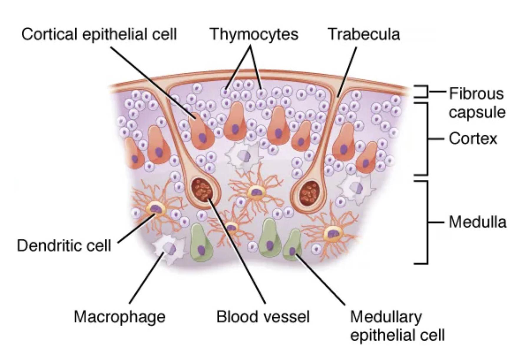
Labeled Components of the Thymus Structure
Cortical epithelial cell: These cells form a supportive framework in the outer layer of the thymus, nurturing developing thymocytes through direct interaction and secretion of signaling molecules. They play a key role in positive selection, ensuring only functional T-cells proceed to maturity.
Thymocytes: As precursors to T-cells, thymocytes undergo rigorous selection processes within the thymus to eliminate those that might attack the body’s own tissues. Their maturation involves stages from double-positive to single-positive cells, critical for immune competence.
Trabecula: Trabeculae are connective tissue septa that divide the thymus into lobules, providing structural support and pathways for blood vessels and nerves. They help maintain the organ’s architecture, facilitating efficient cell migration and interaction.
Fibrous capsule: This outer layer encases the entire thymus, offering protection and separating it from surrounding tissues. Composed of dense connective tissue, it anchors the gland and allows for expansion during early life when the thymus is most active.
Cortex: The cortex is the outer region densely packed with thymocytes, where initial stages of T-cell development occur. It features a high concentration of epithelial cells and is vital for the proliferation and early selection of immature T-cells.
Dendritic cell: Dendritic cells in the thymus present antigens to thymocytes, aiding in negative selection to remove self-reactive cells. Their branched morphology enhances antigen capture and presentation, ensuring immune tolerance.
Medulla: Situated centrally, the medulla contains fewer thymocytes but more mature T-cells ready for export. It hosts Hassall’s corpuscles, which may assist in regulatory T-cell development and final maturation steps.
Macrophage: Macrophages patrol the thymus, phagocytosing apoptotic cells and debris from failed thymocytes. They contribute to the clean-up process, maintaining a healthy environment for ongoing T-cell selection.
Blood vessel: Blood vessels supply nutrients and oxygen to the thymus while facilitating the entry of precursor cells and exit of mature T-cells. They form a network that supports the organ’s metabolic demands and immune cell trafficking.
Medullary epithelial cell: These cells in the medulla express self-antigens via AIRE gene regulation, crucial for central tolerance. They interact with thymocytes to induce apoptosis in those recognizing self-antigens, preventing autoimmunity.
The Anatomy of the Thymus Gland
The thymus is a bilobed organ that reaches its peak size during puberty before gradually involuting with age. Understanding its microscopic structure reveals how it orchestrates immune education.
-
-
- The fibrous capsule surrounds the gland, dividing it into lobes via trabeculae that extend inward.
- Each lobe consists of an outer cortex and inner medulla, creating distinct microenvironments for T-cell development.
- Cortical epithelial cells line the cortex, forming a reticular network that supports thymocyte proliferation.
- In the medulla, medullary epithelial cells and Hassall’s corpuscles provide a site for final checks on T-cell reactivity.
- Blood vessels penetrate through trabeculae, ensuring efficient delivery of hematopoietic precursors from bone marrow.
- Dendritic cells and macrophages are interspersed, acting as sentinels for antigen presentation and waste removal.
-
This sectional view highlights the trabecular extensions, illustrating how they compartmentalize the thymus for optimized function.
Physiological Role of the Thymus in Immunity
The thymus is indispensable for generating a diverse T-cell repertoire, which underpins cellular immunity against viruses and cancers. Its activity peaks in childhood, producing millions of T-cells daily to populate peripheral lymphoid organs.
-
-
- Thymocytes enter the cortex as double-negative cells, progressing to double-positive stages under epithelial cell guidance.
- Positive selection in the cortex ensures T-cells can recognize MHC molecules, with non-functional cells undergoing apoptosis.
- Surviving thymocytes migrate to the medulla for negative selection, where dendritic cells and medullary epithelial cells present self-antigens.
- Macrophages clear apoptotic debris, preventing inflammation and maintaining thymic homeostasis.
- Mature single-positive T-cells exit via blood vessels, entering circulation to patrol for threats.
- Hormones like thymosin and thymopoietin, secreted by epithelial cells, regulate thymocyte differentiation and proliferation.
-
Age-related thymic involution reduces output, contributing to immunosenescence, but residual function persists into adulthood.
Cellular Interactions Within the Thymus
Interactions between stromal cells and thymocytes are mediated by cytokines, chemokines, and cell adhesion molecules. These dynamics ensure precise control over T-cell fate decisions.
-
-
- Cortical epithelial cells express IL-7, promoting thymocyte survival and expansion in early stages.
- Dendritic cells, derived from bone marrow, cluster in the cortico-medullary junction for efficient antigen sampling.
- Medullary epithelial cells, through AIRE expression, promote ectopic gene transcription, exposing thymocytes to tissue-specific antigens.
- Macrophages not only phagocytose but also secrete cytokines that modulate the thymic microenvironment.
- Trabeculae and the fibrous capsule provide mechanical support, while allowing neural innervation that influences thymic function.
- Blood vessels maintain a barrier, preventing premature antigen exposure that could disrupt tolerance induction.
-
This intricate cellular dialogue underscores the thymus’s role as an immune educator.
Developmental Stages and Thymic Function
Thymic development begins in utero, with epithelial-mesenchymal interactions forming the organ’s structure. Postnatally, it adapts to environmental cues to fine-tune immune responses.
-
-
- Precursor cells from bone marrow seed the thymus, differentiating under stromal influence.
- The cortex supports rapid proliferation, with thymocytes undergoing V(D)J recombination for TCR diversity.
- In the medulla, regulatory T-cells develop, crucial for suppressing autoimmunity.
- Epithelial cells in both regions secrete factors like T3 and T4? Wait, no?actually, the thymus does not produce thyroid hormones; that’s the thyroid gland. Instead, it releases thymic hormones such as thymulin.
- Dendritic cells and macrophages ensure quality control, eliminating up to 95% of thymocytes.
- Mature T-cells emigrate, contributing to naive T-cell pools in spleen and lymph nodes.
-
Understanding these stages highlights the thymus’s pivotal position in immunity.
The thymus, though often overlooked, is foundational to a robust immune system, adapting throughout life to maintain balance between protection and tolerance. Its structural elegance, as depicted in this sectional view, mirrors the complexity of T-cell maturation, ensuring lifelong immunological vigilance.
- Research explores thymic regeneration for enhancing immunity in the elderly or immunocompromised individuals.
In summary, the thymus stands as a cornerstone of adaptive immunity, with its intricate structure and histology underpinning lifelong protection against diseases. Appreciating its location and interactions with other organs highlights the elegance of the human immune system.

