The thymus gland plays a crucial role in the development of the immune system, serving as the primary site for T-cell maturation. Located in the upper chest, just above the heart, this organ is essential for adaptive immunity, ensuring the body can effectively combat pathogens and maintain health throughout life.
Key Labeled Structures in the Image
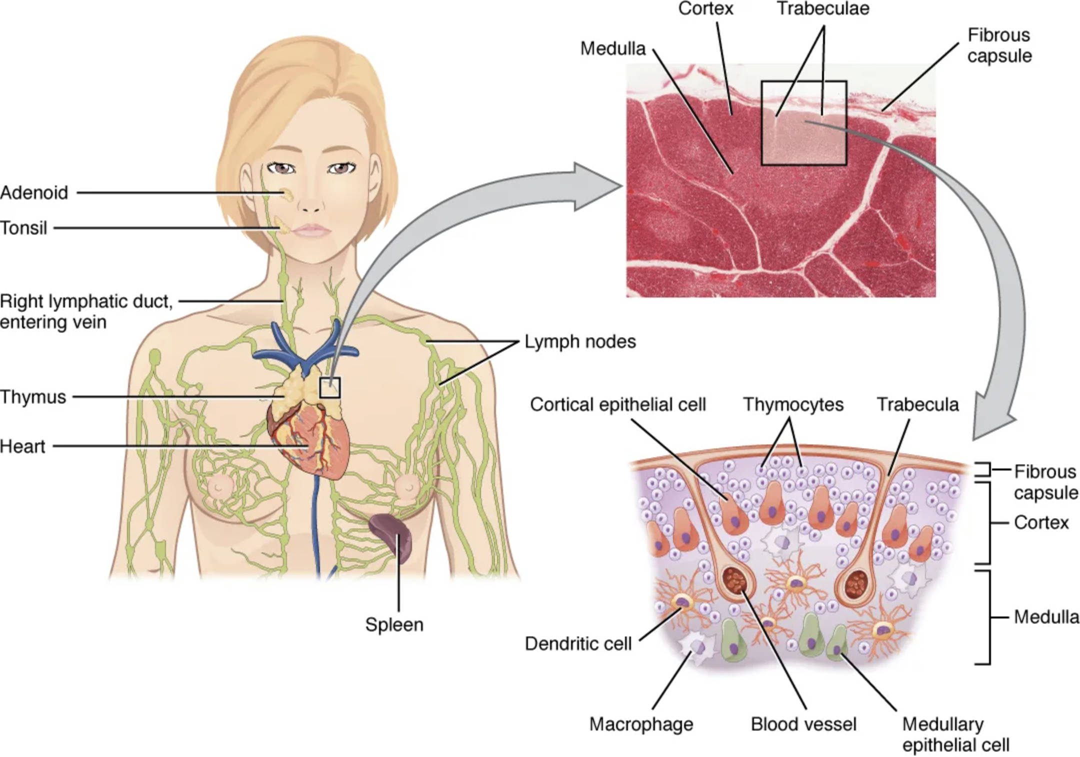
Adenoid The adenoid, also known as the pharyngeal tonsil, is a mass of lymphoid tissue located at the back of the nasal cavity. It acts as a first line of defense against inhaled pathogens by trapping and destroying them through immune responses.
Tonsil The tonsil refers to the palatine tonsils, which are lymphoid organs situated on either side of the throat. They help protect against infections entering through the mouth by producing antibodies and facilitating immune cell activation.
Right lymphatic duct, entering vein The right lymphatic duct collects lymph from the right side of the body and drains it into the venous system via the right subclavian vein. This structure ensures the return of lymph fluid to the bloodstream, maintaining fluid balance and transporting immune cells.
Thymus The thymus is a bilobed gland located in the mediastinum, superior to the heart. It is vital for the maturation of T-lymphocytes, which are key players in cell-mediated immunity.
Heart The heart is the central organ of the cardiovascular system, pumping blood throughout the body. In this context, it serves as a landmark for the position of the thymus, which lies directly above it.
Spleen The spleen is an abdominal organ involved in filtering blood and recycling old red blood cells. It also functions as a secondary lymphoid organ, supporting immune responses against blood-borne pathogens.
Lymph nodes Lymph nodes are small, bean-shaped structures distributed along lymphatic vessels. They filter lymph fluid, trap antigens, and provide sites for immune cell interactions to initiate responses.
Cortex The cortex is the outer layer of lymphoid organs like the thymus and lymph nodes, densely packed with lymphocytes. In the thymus, it is where immature T-cells proliferate and begin their selection process.
Medulla The medulla is the inner region of the thymus and other lymphoid organs, appearing lighter in staining due to fewer cells. It contains mature T-cells and epithelial cells that aid in the final stages of T-cell education.
Trabeculae Trabeculae are connective tissue septa that divide the thymus into lobules. They provide structural support and house blood vessels that supply nutrients to the gland.
Fibrous capsule The fibrous capsule is a tough outer covering of connective tissue surrounding organs like the thymus and spleen. It protects the internal structures and allows for expansion during immune activity.
Cortical epithelial cell The cortical epithelial cell forms part of the thymic stroma in the cortex. These cells nurture developing thymocytes and present self-antigens to ensure proper T-cell selection.
Thymocytes Thymocytes are precursor cells to mature T-lymphocytes found primarily in the thymus. They undergo rigorous selection processes to become immunocompetent while avoiding autoimmunity.
Trabecula A trabecula is a single strand of the trabeculae framework within the thymus. It separates lobules and facilitates the organization of thymic tissue.
Dendritic cell The dendritic cell is an antigen-presenting cell found in lymphoid tissues like the spleen. It captures antigens and presents them to T-cells, initiating adaptive immune responses.
Macrophage The macrophage is a phagocytic cell that engulfs and digests pathogens and debris in organs such as the spleen. It also presents antigens and secretes cytokines to modulate immunity.
Blood vessel The blood vessel in lymphoid organs transports blood, delivering oxygen and nutrients while removing waste. In the spleen, it allows for the filtration of blood components.
Medullary epithelial cell The medullary epithelial cell resides in the medulla of the thymus. It expresses tissue-specific antigens to promote central tolerance in maturing T-cells.
The Anatomical Location of the Thymus
The thymus is strategically positioned in the body to support immune development from an early age. Understanding its placement relative to other structures enhances appreciation of its function in overall health.
- Situated in the anterior superior mediastinum, the thymus lies behind the sternum and anterior to the great vessels.
- It extends from the lower border of the thyroid gland down to the fourth costal cartilage.
- In infants and children, the gland is relatively large, but it undergoes involution with age, gradually being replaced by fatty tissue.
- Proximity to the heart and major veins allows efficient circulation of immune cells.
- Lymphatic vessels connect it to surrounding nodes, facilitating lymph drainage and immune surveillance.
Structural Organization and Histology
The structure of the thymus is uniquely adapted for its role in T-cell maturation. Histological features reveal distinct zones that support different stages of immune cell development.
- Encased in a thin fibrous capsule, the thymus is divided into multiple lobules by trabeculae extending inward.
- Each lobule consists of a peripheral cortex and a central medulla, visible in micrographs as darker and lighter areas respectively.
- The cortex is rich in densely packed thymocytes, interspersed with epithelial cells and macrophages.
- In the medulla, Hassall’s corpuscles—concentric layers of epithelial cells—aid in the removal of apoptotic cells.
- Blood-thymus barrier in the cortex prevents premature antigen exposure, ensuring proper T-cell education.
- Histologically, the thymus stains with hematoxylin and eosin, highlighting cellular density variations.
Physiological Functions of the Thymus
The thymus is indispensable for establishing a competent immune system. It produces hormones that influence immune cell development beyond its structural role.
- Primarily, it facilitates the differentiation of bone marrow-derived precursors into mature T-cells.
- Positive and negative selection processes in the cortex and medulla eliminate self-reactive cells, preventing autoimmunity.
- Thymic hormones like thymosin and thymopoietin stimulate T-cell proliferation and maturation.
- The gland’s output peaks during childhood, contributing to lifelong immunity.
- It interacts with the endocrine system, responding to stress hormones that can temporarily suppress its function.
- Beyond T-cells, it influences natural killer cell activity and overall immune homeostasis.
Related Lymphoid Organs and Their Interactions
The thymus does not operate in isolation but integrates with other lymphoid structures. This interconnectedness ensures comprehensive immune protection across the body.
- Lymph nodes, scattered throughout, act as filters where antigens meet immune cells.
- The spleen complements the thymus by handling blood-borne threats, with its white pulp areas mimicking lymph node architecture.
- Tonsils and adenoids form the Waldeyer’s ring, guarding entry points against respiratory and oral pathogens.
- Lymphatic ducts, such as the right lymphatic duct, return filtered lymph to circulation, distributing mature T-cells systemically.
- These organs collectively maintain vigilance, with the thymus providing the educated T-cells essential for coordination.
Clinical Significance and Age-Related Changes
Awareness of thymic changes over time is vital for understanding immune health. Alterations in structure can impact disease susceptibility and treatment approaches.
- In neonates, the thymus is at its largest relative size, supporting rapid immune system establishment.
- Age-related involution begins post-puberty, reducing active tissue but not entirely abolishing function.
- Conditions like myasthenia gravis involve thymic abnormalities, often requiring imaging for diagnosis.
- Thymectomy, surgical removal, is performed in certain autoimmune disorders, with minimal long-term effects if done after maturity.
- Research explores thymic regeneration for enhancing immunity in the elderly or immunocompromised individuals.
In summary, the thymus stands as a cornerstone of adaptive immunity, with its intricate structure and histology underpinning lifelong protection against diseases. Appreciating its location and interactions with other organs highlights the elegance of the human immune system.

